World J Mens Health.
2018 Jan;36(1):66-72. 10.5534/wjmh.17025.
Feasibility of Polycaprolactone Scaffolds Fabricated by Three-Dimensional Printing for Tissue Engineering of Tunica Albuginea
- Affiliations
-
- 1Department of Urology, Chonnam National University Medical School, Chonnam National University Sexual Medicine Research Center, Gwangju, Korea. uropark@gmail.com
- 2Nano Convergence and Manufacturing Systems Research Division, Korea Institute of Machinery and Materials (KIMM), Daejeon, Korea.
- 3MEMS and Nanotechnology Laboratory, School of Mechanical Systems Engineering, Chonnam National University, Gwangju, Korea.
- KMID: 2399173
- DOI: http://doi.org/10.5534/wjmh.17025
Abstract
- PURPOSE
To investigate the feasibility of a polycaprolactone (PCL) scaffold fabricated by three-dimensional (3D) printing for tissue engineering applications for tunica albuginea.
MATERIALS AND METHODS
PCL scaffolds were fabricated by use of a 3D printing system. Two scaffolds were fabricated that differed in the architecture of the lay-down pattern: a 90°PCL scaffold and a 45°PCL scaffold. Mechanical properties were measured to compare tensile strength between the two scaffold types. The scaffolds were characterized by scanning electron microscope (SEM) images. The scaffolds were seeded with fibroblast cells, and the ability of these scaffolds to support the cells was evaluated by immunofluorescence staining.
RESULTS
The PCL scaffolds had well-structured shapes, regular arrays, and good interconnection in SEM images. The horizontal and vertical Young's modulus coefficients were 13 and 12 MPa for the 90°PCL scaffold and 19 and 21 MPa for the 45°PCL scaffold, respectively. Microscopy images revealed that human fibroblast cells covered the entire scaffold surface. Immunofluorescence staining of ER-TR7 confirmed that the fibroblast cells remained viable and proliferated throughout the time course of the culture.
CONCLUSIONS
This preliminary study provides experimental evidence for the feasibility of 3D printing of PCL scaffolds for tissue engineering applications of tunica albuginea.
MeSH Terms
Figure
-
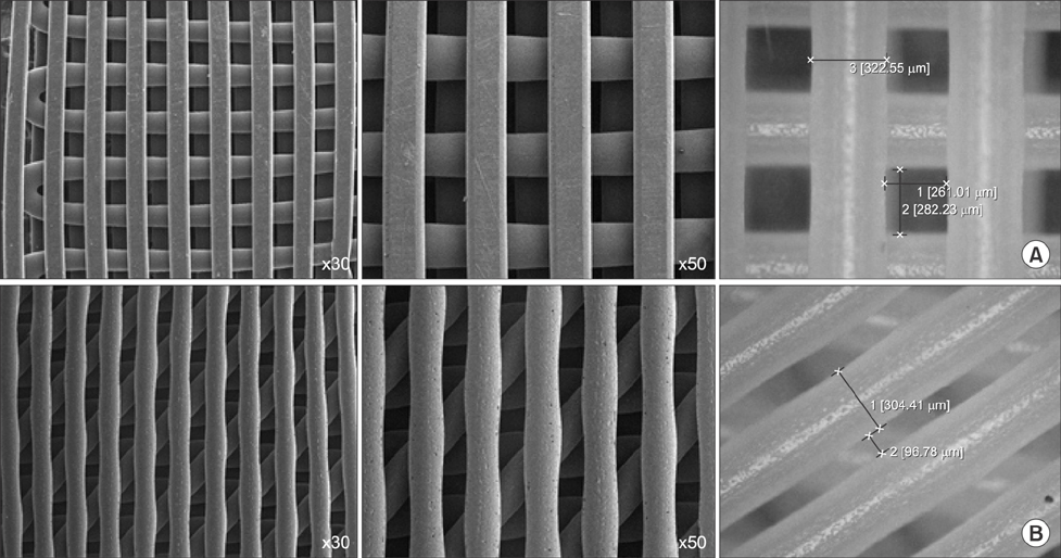
Fig. 1 Scanning electron micrograph images of 90° plotted polycaprolactone (PCL) 300/300 scaffold (A) and 45° plotted PCL 300/300 scaffold (B).
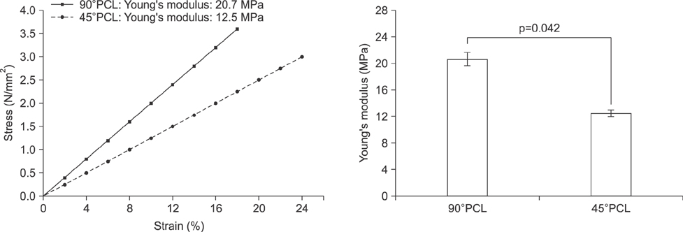
Fig. 2 Mechanical properties of three-dimensional printed polycaprolactone (PCL) scaffolds. Mean tensile stress, strain, and Young's modulus values of two different types of PCL scaffolds.
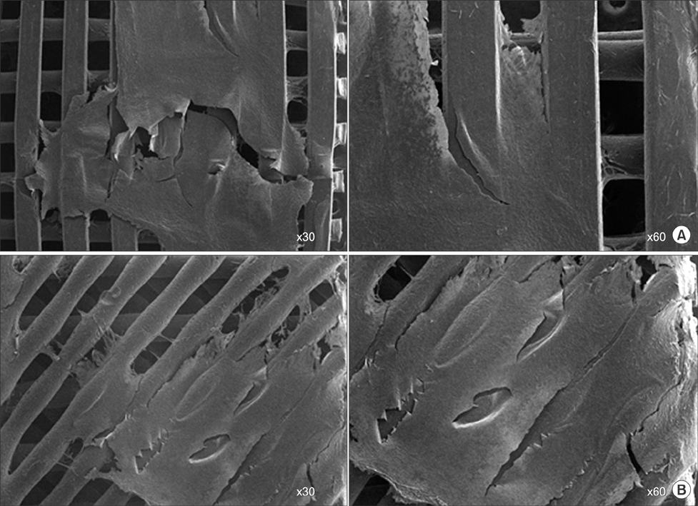
Fig. 3 Scanning electron microscopic images of fibroblasts on 90° polycaprolactone (PCL) scaffold (A) and 45°PCL scaffold cultured for 2 weeks in vitro (B).

Fig. 4 Immunofluorescence staining of fibroblast cells with DAPI (4',6-diamidino-2-phenylindole) and ER-TR7 on 90° (A) and 45° (A, B) polycaprolactone (PCL) scaffolds. Confocal microscopic images of 45°PCL scaffolds (B).
Reference
-
1. Garriboli M, Radford A, Southgate J. Regenerative medicine in urology. Eur J Pediatr Surg. 2014; 24:227–236.
Article2. Osman NI, Hillary C, Bullock AJ, MacNeil S, Chapple CR. Tissue engineered buccal mucosa for urethroplasty: progress and future directions. Adv Drug Deliv Rev. 2015; 82-83:69–76.
Article3. Takeuchi M, Masumori N, Tsukamoto T. Ureteral reconstruction with bowel segments: experience with eight patients in a single institute. Korean J Urol. 2014; 55:742–749.
Article4. Lam Van Ba O, Aharony S, Loutochin O, Corcos J. Bladder tissue engineering: a literature review. Adv Drug Deliv Rev. 2015; 82-83:31–37.
Article5. Kim JH, Lee HJ, Song YS. Treatment of bladder dysfunction using stem cell or tissue engineering technique. Korean J Urol. 2014; 55:228–238.
Article6. Damaser MS, Sievert KD. Tissue engineering and regenerative medicine: bench to bedside in urology. Preface. Adv Drug Deliv Rev. 2015; 82-83:v–vii.7. Zhang C, Murphy SV, Atala A. Regenerative medicine in urology. Semin Pediatr Surg. 2014; 23:106–111.
Article8. Wefer J, Schlote N, Sekido N, Sievert KD, Wefer AE, Nunes L, et al. Tunica albuginea acellular matrix graft for penile reconstruction in the rabbit: a model for treating Peyronie's disease. BJU Int. 2002; 90:326–331.
Article9. Schultheiss D, Lorenz RR, Meister R, Westphal M, Gabouev AI, Mertsching H, et al. Functional tissue engineering of autologous tunica albuginea: a possible graft for Peyronie's disease surgery. Eur Urol. 2004; 45:781–786.
Article10. Imbeault A, Bernard G, Ouellet G, Bouhout S, Carrier S, Bolduc S. Surgical option for the correction of Peyronie's disease: an autologous tissue-engineered endothelialized graft. J Sex Med. 2011; 8:3227–3235.
Article11. Ledger WJ. Current problems in antibiotic treatment in obstetrics and gynecology. Rev Infect Dis. 1985; 7:Suppl 4. S679–S689.
Article12. Cipitri A, Skelton A, Dargaville TR, Dalton PD, Hutmacher DW. Design, fabrication and characterization of PCL electrospun scaffolds-a review. J Mater Chem. 2011; 21:9419–9453.13. Park SA, Kim HJ, Lee SH, Lee JH, Kim HK, Yoon TR, et al. Fabrication of nano/microfiber scaffolds using a combination of rapid prototyping and electrospinning systems. Polym Eng Sci. 2011; 51:1883–1890.
Article14. Brock G, Hsu GL, Nunes L, von Heyden B, Lue TF. The anatomy of the tunica albuginea in the normal penis and Peyronie's disease. J Urol. 1997; 157:276–281.
Article15. Hollister SJ. Porous scaffold design for tissue engineering. Nat Mater. 2005; 4:518–524.
Article16. Chia HN, Wu BM. Recent advances in 3D printing of biomaterials. J Biol Eng. 2015; 9:4.
Article17. Ferretti L, Giuliani M, Bessède T, Qiu X, Zhang H, Alsaid B, et al. Tissue engineering for penile surgery: comparative study of noncellular and cell-seeded synthetic grafts for tunica albuginea replacement. J Sex Med. 2012; 9:625–631.
Article18. Lue TF. Physiology of penile erection and pathophysiology of erectile dysfunction. In : Wein AJ, Kavoussi LR, Partin AW, Peters CA, editors. Campbell-Walsh urology. 11th ed. Philadelphia: Elsevier;2016. p. 612–642.
- Full Text Links
-
- Actions
-
Cited
- CITED
-
- Close
- Share
-
- Similar articles
-
- Solid Freeform Techniques Application in Bone Tissue Engineering for Scaffold Fabrication
- Current Advances in Three-Dimensional Tissue/Organ Printing
- Current Status of Three-Dimensional Printing Inks for Soft Tissue Regeneration
- Electrospinning Fabrication Methods to Incorporate Laminin in Polycaprolactone for Kidney Tissue Engineering
- Three-Dimensional Printing in Tissue Engineering and Regenerative Medicine
