A rare case of multiple pituitary adenomas in an adolescent Cushing disease presenting as a vertebral compression fracture
- Affiliations
-
- 1Department of Pediatrics, Pusan National University Children’s Hospital, Yangsan, Korea. ymk@pusan.ac.kr
- 2Department of Otorhinolaryngology, Pusan National University Yangsan Hospital, Yangsan, Korea.
- 3Department of Neurosergery, Pusan National University Yangsan Hospital, Yangsan, Korea.
- 4Department of Radiology, Pusan National University Yangsan Hospital, Yangsan, Korea.
- 5Department of Pathology, Pusan National University Yangsan Hospital, Yangsan, Korea.
- KMID: 2396140
- DOI: http://doi.org/10.6065/apem.2017.22.3.197
Abstract
- Cushing disease in children and adolescents, especially with multiple pituitary adenomas (MPAs), is very rare. We report 17-year-old boy with MPAs. He presented with a vertebral compression fracture, weight gain, short stature, headache, and hypertension. On magnetic resonance imaging (MRI), only a left pituitary microadenoma was found. After surgery, transient clinical improvement was observed but headache and hypertension were observed again after 3 months later. Follow-up MRI showed a newly developed right pituitary microadenoma 6 months after the surgery. The need for careful clinical and radiographic follow-up should be emphasized in the search for potential MPAs in patients with persistent Cushing disease.
MeSH Terms
Figure
-
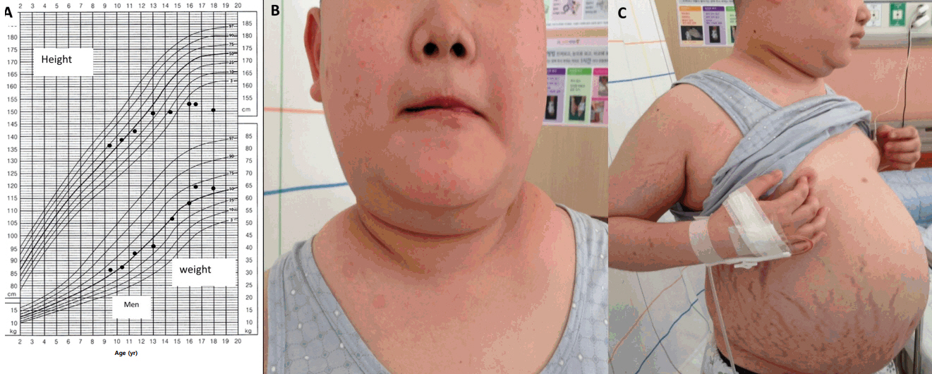
Fig. 1. Growth curve (A) and clinical photos of a patient with Cushing disease showing typical Cushingoid features, including a moon shaped face, buffalo hump, multiple abdominal striae, central obesity, and hypertrichosis (B, C).
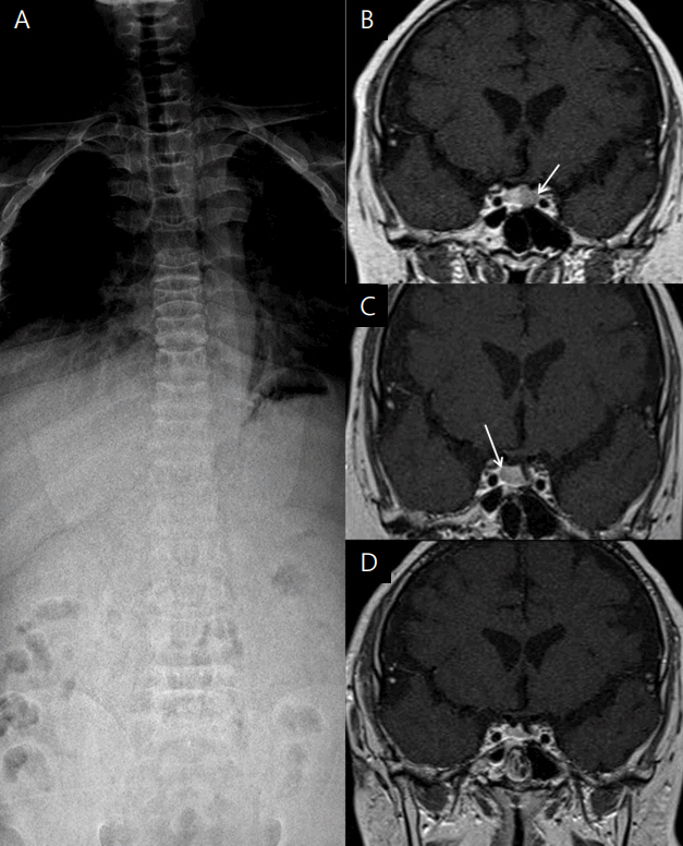
Fig. 2. Simple spinal X-ray and serial pituitary magnetic resonance images of a patient with Cushing disease. (A) Spinal X-ray showing diffuse osteopenia and a compression fracture of the T9, L2, and T3 vertebral bodies. (B) T2-weighted coronal view showing an 8-mm microadenoma in the left pituitary gland (white arrow). (C) T2-weighted coronal view after the first operation showing a 9-mm microadenoma in the right pituitary gland (white arrow). (D) T2-weighted coronal view after the second operation showing a homogenous pituitary gland.
Reference
-
References
1. Lacroix A, Feelders RA, Stratakis CA, Nieman LK. Cushing's syndrome. Lancet. 2015; 386:913–27.
Article2. Storr HL, Chan LF, Grossman AB, Savage MO. Paediatric Cushing's syndrome: epidemiology, investigation and therapeutic advances. Trends Endocrinol Metab. 2007; 18:167–74.
Article3. Stratakis CA. Cushing syndrome in pediatrics. Endocrinol Metab Clin North Am. 2012; 41:793–803.
Article4. Storr HL, Savage MO. Management of endocrine disease: paediatric Cushing's disease. Eur J Endocrinol. 2015; 173:R35–45.
Article5. Güemes M, Murray PG, Brain CE, Spoudeas HA, Peters CJ, Hindmarsh PC, et al. Management of Cushing syndrome in children and adolescents: experience of a single tertiary centre. Eur J Pediatr. 2016; 175:967–76.
Article6. Storr HL, Drake WM, Evanson J, Matson M, Berney DM, Grossman AB, et al. Endonasal endoscopic transsphenoidal pituitary surgery: early experience and outcome in paediatric Cushing's disease. Clin Endocrinol (Oxf). 2014; 80:270–6.7. Petersenn S, Beckers A, Ferone D, van der Lely A, Bollerslev J, Boscaro M, et al. Therapy of endocrine disease: outcomes in patients with Cushing's disease undergoing transsphenoidal surgery: systematic review assessing criteria used to define remission and recurrence. Eur J Endocrinol. 2015; 172:R227–39.
Article8. Pivonello R, De Leo M, Cozzolino A, Colao A. The treatment of Cushing's disease. Endocr Rev. 2015; 36:385–486.
Article9. Zampetti B, Grossrubatscher E, Dalino Ciaramella P, Boccardi E, Loli P. Bilateral inferior petrosal sinus sampling. Endocr Connect. 2016; 5:R12–25.
Article10. Sheth SA, Mian MK, Neal J, Tritos NA, Nachtigall L, Klibanski A, et al. Transsphenoidal surgery for cushing disease after nondiagnostic inferior petrosal sinus sampling. Neurosurgery. 2012; 71:14–22.
Article11. Potts MB, Shah JK, Molinaro AM, Blevins LS, Tyrrell JB, Kunwar S, et al. Cavernous and inferior petrosal sinus sampling and dynamic magnetic resonance imaging in the preoperative evaluation of Cushing's disease. J Neurooncol. 2014; 116:593–600.
Article12. Budan RM, Georgescu CE. Multiple pituitary adenomas: a systematic review. Front Endocrinol (Lausanne). 2016; 7:1.
Article13. Pu J, Wang Z, Zhou H, Zhong A, Jin K, Ruan L, et al. Isolated double adrenocorticotropic hormone-secreting pituitary adenomas: a case report and review of the literature. Oncol Lett. 2016; 12:585–90.
Article14. Rutkowski MJ, Flanigan PM, Aghi MK. Update on the management of recurrent Cushing's disease. Neurosurg Focus. 2015; 38:E16.
Article15. Bertagna X, Guignat L. Approach to the Cushing's disease patient with persistent/recurrent hypercortisolism after pituitary surgery. J Clin Endocrinol Metab. 2013; 98:1307–18.
Article16. Chandler WF, Barkan AL, Hollon T, Sakharova A, Sack J, Brahma B, et al. Outcome of transsphenoidal surgery for Cushing disease: a single-center experience over 32 years. Neurosurgery. 2016; 78:216–23.17. Lonser RR, Ksendzovsky A, Wind JJ, Vortmeyer AO, Oldfield EH. Prospective evaluation of the characteristics and incidence of adenoma-associated dural invasion in Cushing disease. J Neurosurg. 2012; 116:272–9.
Article18. Savage MO, Chan LF, Afshar F, Plowman PN, Grossman AB, Storr HL. Advances in the management of paediatric Cushing's disease. Horm Res. 2008; 69:327–33.
Article
- Full Text Links
-
- Actions
-
Cited
- CITED
-
- Close
- Share
-
- Similar articles
-
- Continuous Multiple Vertebral Compression Fractures in Multiple Myeloma Patient
- Pregnancy and Lactation-associated Osteoporosis with Vertebral Compression Fracture
- Unsuspected Multiple Myeloma Diagnosed by Bone Biopsy during Percutaneous Kyphoplasty in Vertebral Compression Fracture
- Intraspinal Extradural Cyst Subsequent to a Vertebral Compression Fracture - A Case Report -
- Recent Progress in the Medical Therapy of Pituitary Tumors
