Magnetic Resonance Imaging (MRI) of a Hypertrophy of Cartilage and Simultaneous Regeneration of a Damaged Meniscus after Autologous Bone Marrow Aspirates Concentrate (BMAC) Transplantation: a Case Report and Literature Review
- Affiliations
-
- 1Department of Radiology, Soonchunhyang University Hospital, Seoul, Korea. reonora@schmc.ac.kr
- KMID: 2392691
- DOI: http://doi.org/10.13104/imri.2017.21.3.187
Abstract
- Bone marrow aspirates concentrate (BMAC) transplantation is a well-known technique for cartilage regeneration with good clinical outcomes for symptoms in patients with osteoarthritis (OA). Magnetic resonance imaging (MRI) has an important role in evaluating the degree of cartilage repair in cartilage regeneration therapy instead of a second assessment via an arthroscopy. We experienced a case of hypertrophic regeneration of the cartilage and a presumed simultaneous regeneration of the posterior horn of the lateral meniscus after BMAC transplantation for a cartilage defect at the lateral tibial and femoral condyle. This report provides the details of a case of an unusual treatment response after a BMAC transplant. This report is the first of its kind to demonstrate a MR image that displays the simultaneous regeneration of the cartilage and meniscus with a differentiation ability of the mesenchymal stem cell to the desired cell lineage.
Keyword
MeSH Terms
Figure
-
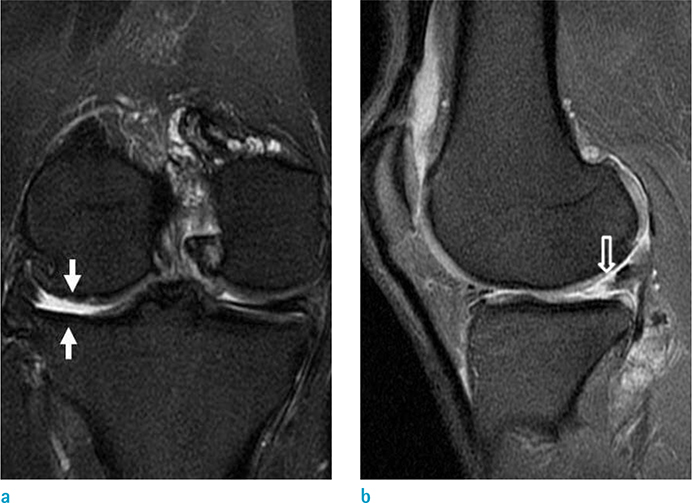
Fig. 1 Right knee MRI of a 44-year-old male presenting with knee pain previously treated at an outside hospital. (a) Coronal fat-saturated T2 weighted image displays high grade cartilage defects (white arrows) in the lateral tibial plateau and lateral femoral condyle. (b) Sagittal fat-saturated proton density weighted image exhibits a complex tear (empty arrow) in the posterior horn of the lateral meniscus.
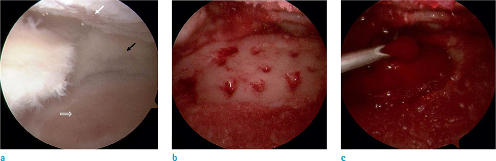
Fig. 2 Arthroscopic finding during the bone marrow aspirates concentrate (BMAC) transplantation. (a) Grade IV cartilage defects at the lateral tibial plateau (empty arrow) and lateral femoral condyle (white arrow) and residual posterior horn of the lateral meniscus (black arrow) after a subtotal meniscectomy are provided. (b) A cartilage defect at the lateral tibial plateau after a microfracture is displayed on an arthroscopic image. (c) BMAC transplantation into the lateral tibial plateau with a scaffold is performed.
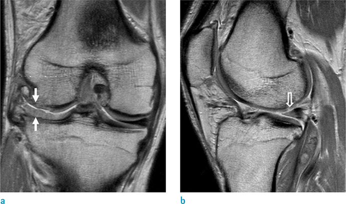
Fig. 3 Follow-up right knee MRI of the patient at 7 weeks after the operation. (a) Coronal proton density weighted image displays hypertrophic regeneration of the cartilage (white arrows) at the lateral tibial plateau and the near complete regeneration of the cartilage at the lateral femoral condyle (white arrows). (b) The sagittal proton density weighted image exhibits a nearly normal-shaped posterior horn of the lateral meniscus (empty arrow) though displays a diffuse increased signal intensity.
Reference
-
1. Chahla J, Dean CS, Moatshe G, Pascual-Garrido C, Serra Cruz R, LaPrade RF. Concentrated bone marrow aspirate for the treatment of chondral injuries and osteoarthritis of the knee: a systematic review of outcomes. Orthop J Sports Med. 2016; 4:2325967115625481.2. Kim YS, Choi YJ, Lee SW, et al. Assessment of clinical and MRI outcomes after mesenchymal stem cell implantation in patients with knee osteoarthritis: a prospective study. Osteoarthritis Cartilage. 2016; 24:237–245.3. Pak J, Lee JH, Park KS, Jeon JH, Lee SH. Potential use of mesenchymal stem cells in human meniscal repair: current insights. Open Access J Sports Med. 2017; 8:33–38.4. Pak J, Lee JH, Lee SH. Regenerative repair of damaged meniscus with autologous adipose tissue-derived stem cells. Biomed Res Int. 2014; 2014:436029.5. Holton J, Imam MA, Snow M. Bone marrow aspirate in the treatment of chondral injuries. Front Surg. 2016; 3:33.6. Marlovits S, Striessnig G, Resinger CT, et al. Definition of pertinent parameters for the evaluation of articular cartilage repair tissue with high-resolution magnetic resonance imaging. Eur J Radiol. 2004; 52:310–319.7. Freitag J, Bates D, Boyd R, et al. Mesenchymal stem cell therapy in the treatment of osteoarthritis: reparative pathways, safety and efficacy - a review. BMC Musculoskelet Disord. 2016; 17:230.8. Chen S, Fu P, Cong R, Wu H, Pei M. Strategies to minimize hypertrophy in cartilage engineering and regeneration. Genes Dis. 2015; 2:76–95.9. Yu H, Adesida AB, Jomha NM. Meniscus repair using mesenchymal stem cells - a comprehensive review. Stem Cell Res Ther. 2015; 6:86.10. Centeno CJ, Busse D, Kisiday J, Keohan C, Freeman M, Karli D. Regeneration of meniscus cartilage in a knee treated with percutaneously implanted autologous mesenchymal stem cells. Med Hypotheses. 2008; 71:900–908.
- Full Text Links
-
- Actions
-
Cited
- CITED
-
- Close
- Share
-
- Similar articles
-
- Bone Marrow Aspiration Concentrate in the Treatment of Osteoarthritis: A Review of its Current Clinical Application
- Bony Union of Osteochondral Lesion of the Talus after Bone Marrow Aspirate Concentrate and Matrix-Induced Chondrogenesis: A Case Report
- Simultaneous Osteoperiosteal Autologous Iliac Crest Graft and Lateral Meniscus Allograft Transplantation for Osteochondral Lesion with Bony Defect and Lateral Discoid Meniscus Tear
- Magnetic resonance imaging Usefulness after Medial Meniscus Posterior Root Tear Repair
- Atelocollagen Scaffold Enhances Cartilage Regeneration in Osteochondral Defects: A Study in Rabbits
