Imaging Sci Dent.
2017 Mar;47(1):17-24. 10.5624/isd.2017.47.1.17.
Detection of furcation involvement using periapical radiography and 2 cone-beam computed tomography imaging protocols with and without a metallic post: An animal study
- Affiliations
-
- 1Department of Stomatology, School of Dentistry, University of São Paulo, São Paulo, Brazil. mgpcaval@usp.br
- KMID: 2391402
- DOI: http://doi.org/10.5624/isd.2017.47.1.17
Abstract
- PURPOSE
The purpose of this study was to assess the accuracy, sensitivity, and specificity of the diagnosis of incipient furcation involvement with periapical radiography (PR) and 2 cone-beam computed tomography (CBCT) imaging protocols, and to test metal artifact interference.
MATERIALS AND METHODS
Mandibular second molars in 10 macerated pig mandibles were divided into those that showed no furcation involvement and those with lesions in the furcation area. Exams using PR and 2 different CBCT imaging protocols were performed with and without a metallic post. Each image was analyzed twice by 2 observers who rated the absence or presence of furcation involvement according to a 5-point scale. Receiver operating characteristic (ROC) curves were used to evaluate the accuracy, sensitivity, and specificity of the observations.
RESULTS
The accuracy of the CBCT imaging protocols ranged from 67.5% to 82.5% in the images obtained with a metallic post and from 72.5% to 80% in those without a metallic post. The accuracy of PR ranged from 37.5% to 55% in the images with a metallic post and from 42.5% to 62.5% in those without a metallic post. The area under the ROC curve values for the CBCT imaging protocols ranged from 0.813 to 0.802, and for PR ranged from 0.503 to 0.448.
CONCLUSION
Both CBCT imaging protocols showed higher accuracy, sensitivity, and specificity than PR in the detection of incipient furcation involvement. Based on these results, CBCT may be considered a reliable tool for detecting incipient furcation involvement following a clinical periodontal exam, even in the presence of a metallic post.
MeSH Terms
Figure
-

Fig. 1 The pig mandibles before (A) and after (B) the chemical lesions were simulated (arrow).
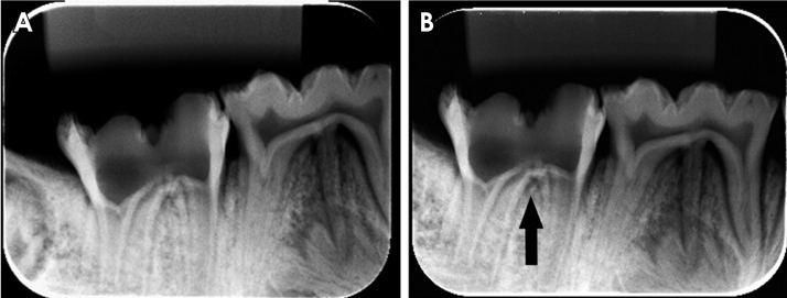
Fig. 2 Periapical radiographs without a lesion (A), and with a lesion (B), illustrating the difficulty in identifying the lesion on periapical radiographs. The lesion is indicated by the arrow in the area where it was created.

Fig. 3 CBCT standard imaging protocol in axial images without a metallic post and without a lesion (A), without a metallic post and with a lesion (B, arrow with brace), with a metallic post and without a lesion (C), and with a metallic post and with a lesion (D, arrow with brace). CBCT, cone-beam computed tomography.
Reference
-
1. American Academy of Periodontology. Glossary of periodontal terms. 4th ed. Chicago: American Academy of Periodontology;2001.2. Hamp SE, Nyman S, Lindhe J. Periodontal treatment of multirooted teeth. Results after 5 years. J Clin Periodontol. 1975; 2:126–135.3. Dannewitz B, Zeidler A, Hüsing J, Saure D, Pfefferle T, Eickholz P, et al. Loss of molars in periodontally treated patients: results 10 years and more after active periodontal therapy. J Clin Periodontol. 2016; 43:53–62.4. Graetz C, Schützhold S, Plaumann A, Kahl M, Springer C, Sälzer S, et al. Prognostic factors for the loss of molars - an 18-years retrospective cohort study. J Clin Periodontol. 2015; 42:943–950.
Article5. Bower RC. Furcation morphology relative to periodontal treatment. Furcation entrance architecture. J Periodontol. 1979; 50:23–27.
Article6. Svärdström G, Wennström JL. Furcation topography of the maxillary and mandibular first molars. J Clin Periodontol. 1988; 15:271–275.
Article7. Mol A. Imaging methods in periodontology. Periodontol 2000. 2004; 34:34–48.
Article8. Cimbaljevic MM, Spin-Neto RR, Miletic VJ, Jankovic SM, Aleksic ZM, Nikolic-Jakoba NS. Clinical and CBCT-based diagnosis of furcation involvement in patients with severe periodontitis. Quintessence Int. 2015; 46:863–870.9. Cavalcanti MG. Cone beam computed tomographic imaging: perspective, challenges, and the impact of near-trend future applications. J Craniofac Surg. 2012; 23:279–282.10. De Vos W, Casselman J, Swennen GR. Cone-beam computerized tomography (CBCT) imaging of the oral and maxillofacial region: a systematic review of the literature. Int J Oral Maxillofac Surg. 2009; 38:609–625.
Article11. Salineiro FC, Pinheiro LR, dos Santos Junior O, Cavalcanti MG. Detection of horizontal root fracture using four different protocols of cone-beam computed tomography. Braz Oral Res. 2015; 29:pii: S1806-83242015000100264.
Article12. Costa FF, Gaia BF, Umetsubo OS, Cavalcanti MG. Detection of horizontal root fracture with small-volume cone-beam computed tomography in the presence and absence of intracanal metallic post. J Endod. 2011; 37:1456–1459.
Article13. Pinheiro LR, Scarfe WC, Augusto de Oliveira Sales M, Gaia BF, Cortes AR, Cavalcanti MG. Effect of cone-beam computed tomography field of view and acquisition frame on the detection of chemically simulated peri-implant bone loss in vitro. J Periodontol. 2015; 86:1159–1165.
Article14. Mohammadpour M, Bakhshalian N, Shahab S, Sadeghi S, Ataee M, Sarikhani S. Effect of titanium and stainless steel posts in detection of vertical root fractures using NewTom VG cone beam computed tomography system. Imaging Sci Dent. 2014; 44:89–94.
Article15. Ferreira RI, Bahrami G, Isidor F, Wenzel A, Haiter-Neto F, Groppo FC. Detection of vertical root fractures by cone-beam computerized tomography in endodontically treated teeth with fiber-resin and titanium posts: an in vitro study. Oral Surg Oral Med Oral Pathol Oral Radiol. 2013; 115:e49–e57.
Article16. Takeshita WM, Iwaki LC, da Silva MC, Sabio S, Albino PR. Comparison of periapical radiography with cone beam computed tomography in the diagnosis of vertical root fractures in teeth with a metallic post. J Conserv Dent. 2014; 17:225–229.17. Santos Junior O, Pinheiro LR, Umetsubo OS, Cavalcanti MG. CBCT-based evaluation of integrity of cortical sinus close to periapical lesions. Braz Oral Res. 2015; 29:pii: S1806-83242015000100216.
Article18. Umetsubo OS, Gaia BF, Costa FF, Cavalcanti MG. Detection of simulated incipient furcation involvement by CBCT: an in vitro study using pig mandibles. Braz Oral Res. 2012; 26:341–347.
Article19. Glickman I. Clinical periodontology: the periodontium in health and disease. 2nd ed. Philadelphia, PA: W.B. Saunders;1958. p. 694–696.20. Landis JR, Koch GG. The measurement of observer agreement for categorical data. Biometrics. 1977; 33:159–174.
Article21. Laky M, Majdalani S, Kapferer I, Frantal S, Gahleitner A, Moritz A, et al. Periodontal probing of dental furcations compared with diagnosis by low-dose computed tomography: a case series. J Periodontol. 2013; 84:1740–1746.
Article22. Walter C, Weiger R, Zitzmann NU. Periodontal surgery in furcation-involved maxillary molars revisited - an introduction of guidelines for comprehensive treatment. Clin Oral Investig. 2011; 15:9–20.23. Bowers GM, Schallhorn RG, McClain PK, Morrison GM, Morgan R, Reynolds MA. Factors influencing the outcome of regenerative therapy in mandibular Class II furcations: Part I. J Periodontol. 2003; 74:1255–1268.
Article24. Štembírek J, Kyllar M, Putnová I, Stehlík L, Buchtová M. The pig as an experimental model for clinical craniofacial research. Lab Anim. 2012; 46:269–279.
Article25. Wang S, Liu Y, Fang D, Shi S. The miniature pig: a useful large animal model for dental and orofacial research. Oral Dis. 2007; 13:530–537.
Article26. Aljehani YA. Diagnostic applications of cone-beam CT for periodontal diseases. Int J Dent. 2014; 2014:865079.
Article27. Costa FF, Gaia BF, Umetsubo OS, Pinheiro LR, Tortamano IP, Cavalcanti MG. Use of large-volume cone-beam computed tomography in identification and localization of horizontal root fracture in the presence and absence of intracanal metallic post. J Endod. 2012; 38:856–859.28. Barbat J, Messer HH. Detectability of artificial periapical lesions using direct digital and conventional radiography. J Endod. 1998; 24:837–842.
Article29. Qiao J, Wang S, Duan J, Zhang Y, Qiu Y, Sun C, et al. The accuracy of cone-beam computed tomography in assessing maxillary molar furcation involvement. J Clin Periodontol. 2014; 41:269–274.
Article30. Costa FF, Pinheiro LR, Umetsubo OS, dos Santos O Jr, Gaia BF, Cavalcanti MG. Influence of cone-beam computed tomographic scan mode for detection of horizontal root fracture. J Endod. 2014; 40:1472–1476.
Article31. Benic GI, Sancho-Puchades M, Jung RE, Deyhle H, Hämmerle CH. In vitro assessment of artifacts induced by titanium dental implants in cone beam computed tomography. Clin Oral Implants Res. 2013; 24:378–383.
- Full Text Links
-
- Actions
-
Cited
- CITED
-
- Close
- Share
-
- Similar articles
-
- Detection of maxillary second molar with two palatal roots using cone beam computed tomography: a case report
- A comparative study of cone-beam computed tomography and digital periapical radiography in detecting mandibular molars root perforations
- Comparison of conventional imaging techniques and CBCT for periodontal evaluation: A systematic review
- Three-dimensional imaging modalities in endodontics
- Cone-beam computed tomography versus digital periapical radiography in the detection of artificially created periapical lesions: A pilot study of the diagnostic accuracy of endodontists using both techniques
