Imaging Sci Dent.
2017 Mar;47(1):25-31. 10.5624/isd.2017.47.1.25.
Cone-beam computed tomography versus digital periapical radiography in the detection of artificially created periapical lesions: A pilot study of the diagnostic accuracy of endodontists using both techniques
- Affiliations
-
- 1Faculty of Dentistry, Estácio de Sá University, Rio de Janeiro, Brazil. fabiovidalmarques@hotmail.com
- 2Faculty of Dentistry, Federal University of Rio de Janeiro, Rio de Janeiro, Brazil.
- KMID: 2391403
- DOI: http://doi.org/10.5624/isd.2017.47.1.25
Abstract
- PURPOSE
The aim of this study was to compare the diagnostic accuracy of previously trained endodontists in the detection of artificially created periapical lesions using cone-beam computed tomography (CBCT) and digital periapical radiography (DPR).
MATERIALS AND METHODS
An ex vivo model using dry skulls was used, in which simulated apical lesions were created and then progressively enlarged using #1/2, #2, #4, and #6 round burs. A total of 11 teeth were included in the study, and 110 images were obtained with CBCT and with an intraoral digital periapical radiographic sensor (Instrumentarium dental, Tuusula, Finland) initially and after each bur was used. Specificity and sensitivity were calculated. All images were evaluated by 10 previously trained, certified endodontists. Agreement was calculated using the kappa coefficient. The accuracy of each method in detecting apical lesions was calculated using the chi-square test.
RESULTS
The kappa coefficient between examiners showed low agreement (range, 0.17-0.64). No statistical difference was found between CBCT and DPR in teeth without apical lesions (P=.15). The accuracy for CBCT was significantly higher than for DPR in all corresponding simulated lesions (P<.001). The correct diagnostic rate for CBCT ranged between 56.9% and 73.6%. The greatest difference between CBCT and DPR was seen in the maxillary teeth (CBCT, 71.4%; DPR, 28.6%; P<.01) and multi-rooted teeth (CBCT, 83.3%; DPR, 33.3%; P<.01).
CONCLUSION
CBCT allowed higher accuracy than DPR in detecting simulated lesions for all simulated lesions tested. Endodontists need to be properly trained in interpreting CBCT scans to achieve higher diagnostic accuracy.
MeSH Terms
Figure
-

Fig. 1 Stabilization guides, positioning tools, X-ray cylinder, and the skull in position for capturing the image.
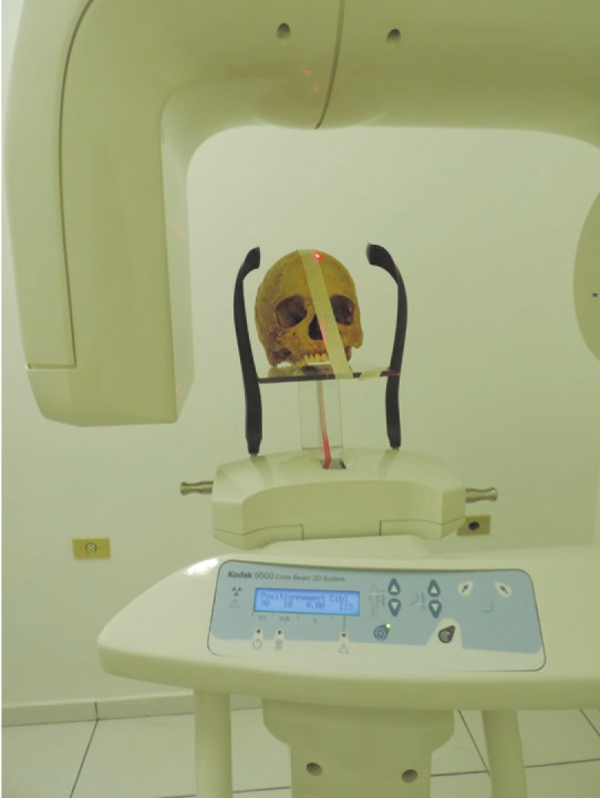
Fig. 2 Acrylic device designed to position the anatomical pieces during cone-beam computed tomography scanning.

Fig. 3 Example of a template with cone-beam computed tomography and digital periapical images evaluated by the examiners.
Reference
-
1. Siqueira JF Jr, Rôças IN, Lopes HP. Treatment of endodontic infections. London: Quintessence;2011.2. Estrela C, Bueno MR, Leles CR, Azevedo B, Azevedo JR. Accuracy of cone beam computed tomography and panoramic and periapical radiography for detection of apical periodontitis. J Endod. 2008; 34:273–279. PMID: 18291274.
Article3. Peters CI, Peters OA. Cone beam computed tomography and other imaging techniques in the determination of periapical healing. Endod Topics. 2012; 26:57–75.
Article4. Folk RB, Thorpe JR, McClanahan SB, Johnson JD, Strother JM. Comparison of two different direct digital radiography systems for the ability to detect artificially prepared periapical lesions. J Endod. 2005; 31:304–306. PMID: 15793390.
Article5. Hadley DL, Replogle KJ, Kirkam JC, Best AM. A comparison of five radiographic systems to D-speed film in the detection of artificial bone lesions. J Endod. 2008; 34:1111–1114. PMID: 18718376.
Article6. White SC, Atchison KA, Hewlett ER, Flack VF. Efficacy of FDA guidelines for prescribing radiographs to detect dental and intraosseous conditions. Oral Surg Oral Med Oral Pathol Oral Radiol Endod. 1995; 80:108–114. PMID: 7552849.
Article7. Patel S, Dawood A, Mannocci F, Wilson R, Pitt Ford T. Detection of periapical bone defects in human jaws using cone bean computed tomography and intraoral radiography. Int Endod J. 2009; 42:507–515. PMID: 19298574.8. Lopez FU, Kopper PM, Cucco C, Della Bona A, de Figueiredo JA, Vier-Pelisser FV. Accuracy of cone-beam computed tomography and periapical radiography in apical periodontitis diagnosis. J Endod. 2014; 40:2057–2060. PMID: 25306306.9. Venskutonis T, Daugela P, Strazdas M, Juodzbalys G. Accuracy of digital radiography and cone beam computed tomography on periapical radiolucency detection in endodontically treated teeth. J Oral Maxillofac Res. 2014; 5:e1.
Article10. Green TL, Walton RE, Taylor JK, Merrell P. Radiographic and histologic periapical findings of root canal treated teeth in cadaver. Oral Surg Oral Med Oral Pathol Oral Radiol Endod. 1997; 83:707–711. PMID: 9195628.
Article11. de Paula-Silva FW, Wu MK, Leonardo MR, da Silva LA, Wesselink PR. Accuracy of periapical radiography and cone-beam computed tomography scans in diagnosing apical periodontitis using histopathological findings as a gold standard. J Endod. 2009; 35:1009–1012. PMID: 19567324.12. Special Committee to revise the joint AAE/AAOMR position statement on use of CBCT in Endodontics. AAE and AAOMR joint position statement: use of cone beam computed tomography in endodontics 2015 update. Oral Surg Oral Med Oral Pathol Oral Radiol. 2015; 120:508–512. PMID: 26346911.13. Tsai P, Torabinejad M, Rice D, Azevedo B. Accuracy of cone-beam computed tomography and periapical radiography in detecting small periapical lesions. J Endod. 2012; 38:965–970. PMID: 22703662.
Article14. Mota de Almeida FJ, Knutsson K, Flygare L. The impact of cone beam computed tomography on the choice of endodontic diagnosis. Int Endod J. 2015; 48:564–572. PMID: 25070420.
Article15. Ganguly R, Ramesh A. Systematic interpretation of CBCT scans: why do it? J Mass Dent Soc. 2014; 62:68–70. PMID: 24624595.16. Soh G, Loh FC, Chong YH. Radiation dosage of a dental imaging system. Quintessence Int. 1993; 24:189–191. PMID: 8511278.17. de Paula-Silva FW, Santamaria M Jr, Leonardo MR, Consolaro A, da Silva LA. Cone-beam computerized tomographic, radiographic, and histologic evaluation of periapical repair in dogs' post-endodontic treatment. Oral Surg Oral Med Oral Pathol Oral Radiol Endod. 2009; 108:796–805. PMID: 19734073.
Article18. Leonardi Dutra K, Haas L, Porporatti AL, Flores-Mir C, Nascimento Santos J, Mezzomo LA, et al. Diagnostic accuracy of cone-bean computed tomography and conventional radiography on apical periodontitis: a systematic review and meta-analysis. J Endod. 2016; 42:356–364. PMID: 26902914.19. Kanagasingam S, Mannocci F, Lim CX, Yong CP, Patel S. Diagnostic accuracy of periapical radiography and cone beam computed tomography in detecting apical periodontitis using histopathological findings as a reference standard. Int Endod J. 2016; (in press).
Article
- Full Text Links
-
- Actions
-
Cited
- CITED
-
- Close
- Share
-
- Similar articles
-
- A comparative study of cone-beam computed tomography and digital periapical radiography in detecting mandibular molars root perforations
- Diagnostic accuracy of artificially induced vertical root fractures: a comparison of direct digital periapical images with conventional periapical images
- Detection of maxillary second molar with two palatal roots using cone beam computed tomography: a case report
- Detection of furcation involvement using periapical radiography and 2 cone-beam computed tomography imaging protocols with and without a metallic post: An animal study
- Detection of peri-implant bone defects using cone-beam computed tomography and digital periapical radiography with parallel and oblique projection
