Anat Cell Biol.
2017 Sep;50(3):239-241. 10.5115/acb.2017.50.3.239.
Unusual muscle of the anterior neck: cadaveric findings with surgical applications
- Affiliations
-
- 1Seattle Science Foundation, Seattle, WA, USA. joei@seattlesciencefoundation.org
- 2Department of Anatomy, Kurume University School of Medicine, Kurume, Japan.
- 3Dental and Oral Medical Center, Kurume University School of Medicine, Kurume, Japan.
- 4Swedish Neuroscience Institute, Swedish Medical Center, Seattle, WA, USA.
- 5Department of Anatomical Sciences, St. George's University, St. George's, Grenada.
- KMID: 2390486
- DOI: http://doi.org/10.5115/acb.2017.50.3.239
Abstract
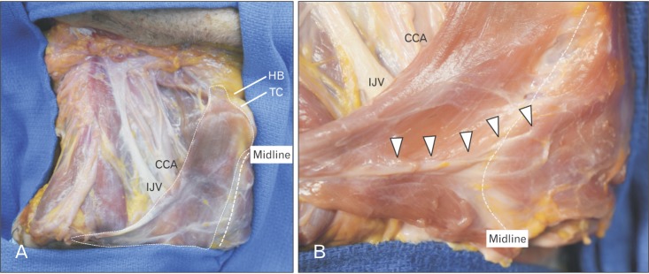
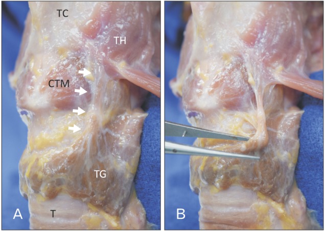
- The omohyoid muscle typically has an inferior belly originating from the superior border of the scapula, and then passes deep to the sternocleidomastoid muscle where its superior belly passes almost vertically upward next to the lateral border of sternohyoid to attach to the inferior border of the body of the hyoid bone lateral to the insertion of sternohyoid. Herein, we report an unusual variant of the omohyoid and sternohyoid muscles. As the omohyoid muscle is commonly used as a surgical landmark during neck dissections, knowledge of its variations such as the one described in the current report is important to surgeons.
Keyword
MeSH Terms
Figure
Reference
-
1. Tubbs RS, Salter EG, Oakes WJ. Unusual origin of the omohyoid muscle. Clin Anat. 2004; 17:578–582. PMID: 15376287.2. Standring S. Gray's anatomy: the anatomical basis of clinical practice. 41st ed. Amsterdam: Elsevier Health Sciences;2015.3. Hatipoğlu ES, Kervancioğlu P, Tuncer MC. An unusual variation of the omohyoid muscle and review of literature. Ann Anat. 2006; 188:469–472. PMID: 16999212.4. Rai R, Ranade A, Nayak S, Vadgaonkar R, Mangala P, Krishnamurthy A. A study of anatomical variability of the omohyoid muscle and its clinical relevance. Clinics (Sao Paulo). 2008; 63:521–524. PMID: 18719765.5. Wood J. Additional varieties in human myology. Proc R Soc Lond. 1865; 14:379–392.6. Tubbs RS, Shoja MM, Loukas M. Bergman's comprehensive encyclopedia of human anatomic variation. Hoboken, NJ: John Wiley & Sons;2016.7. Loth E. Anthropologie des parties molles. Paris: Masson;1931.8. Buntine JA. The omohyoid muscle and fascia: morphology and anomalies. Aust N Z J Surg. 1970; 40:86–88. PMID: 5272615.9. Mizen KD, Mitchell DA. Anatomical variability of omohyoid and its relevance in oropharyngeal cancer. Br J Oral Maxillofac Surg. 2005; 43:285–288. PMID: 15993280.10. Liu SH, Chao KS, Leu YS, Lee JC, Liu CJ, Huang YC, Chang YF, Chen HW, Tsai JT, Chen YJ. Guideline and preliminary clinical practice results for dose specification and target delineation for postoperative radiotherapy for oral cavity cancer. Head Neck. 2015; 37:933–939. PMID: 24634078.
- Full Text Links
- Actions
-
Cited
- CITED
-
- Close
- Share
- Similar articles
-
- Anatomy and variations of digastric muscle
- Normal Anatomy of the Anal Wall and Perianal Spaces: An EUS, MRI and Cadaveric Correlative Study
- The Unusual Origin of the Sternocleidomastoid Artery from the Lingual Artery
- A clinical perspective on the anatomical study of digastric muscle
- A biceps-bicaudatus sartorius muscle: dissection of a variant with possible clinical implications


