A biceps-bicaudatus sartorius muscle: dissection of a variant with possible clinical implications
- Affiliations
-
- 1Department of Anatomy and Surgical Anatomy, School of Medicine, Faculty of Health Sciences, Aristotle University of Thessaloniki, Thessaloniki, Greece
- 2Department of Anatomy, School of Medicine, Faculty of Health Sciences, National and Kapodistrian University of Athens, Athens, Greece
- 3Department of Orthopaedics and Traumatology, 251 Hellenic Air Force General Hospital of Athens, Athens, Greece
- KMID: 2554248
- DOI: http://doi.org/10.5115/acb.23.254
Abstract
- The current cadaveric report describes an unusual morphology of the sartorius muscle (SM), the biceps-bicaudatus variant. The SM had two (lateral and medial) heads, with distinct tendinous origins from the anterior superior iliac spine. The lateral head was further split into a lateral and a medial bundle. The anterior cutaneous branch of the femoral nerve emerged between the origins of the lateral and medial heads. SM morphological variants are exceedingly uncommon, with only a few documented cases in the literature, and several terms used for their description. Although their rare occurrence, they may play an important role in the differential diagnosis of entrapment syndromes, in cases of neural compressions, such as meralgia paresthetica, while careful dissection during the superficial inter-nervous plane of the direct anterior hip approach is of utmost importance, to avoid adverse effects due to the altered SM morphology.
Keyword
Figure
-
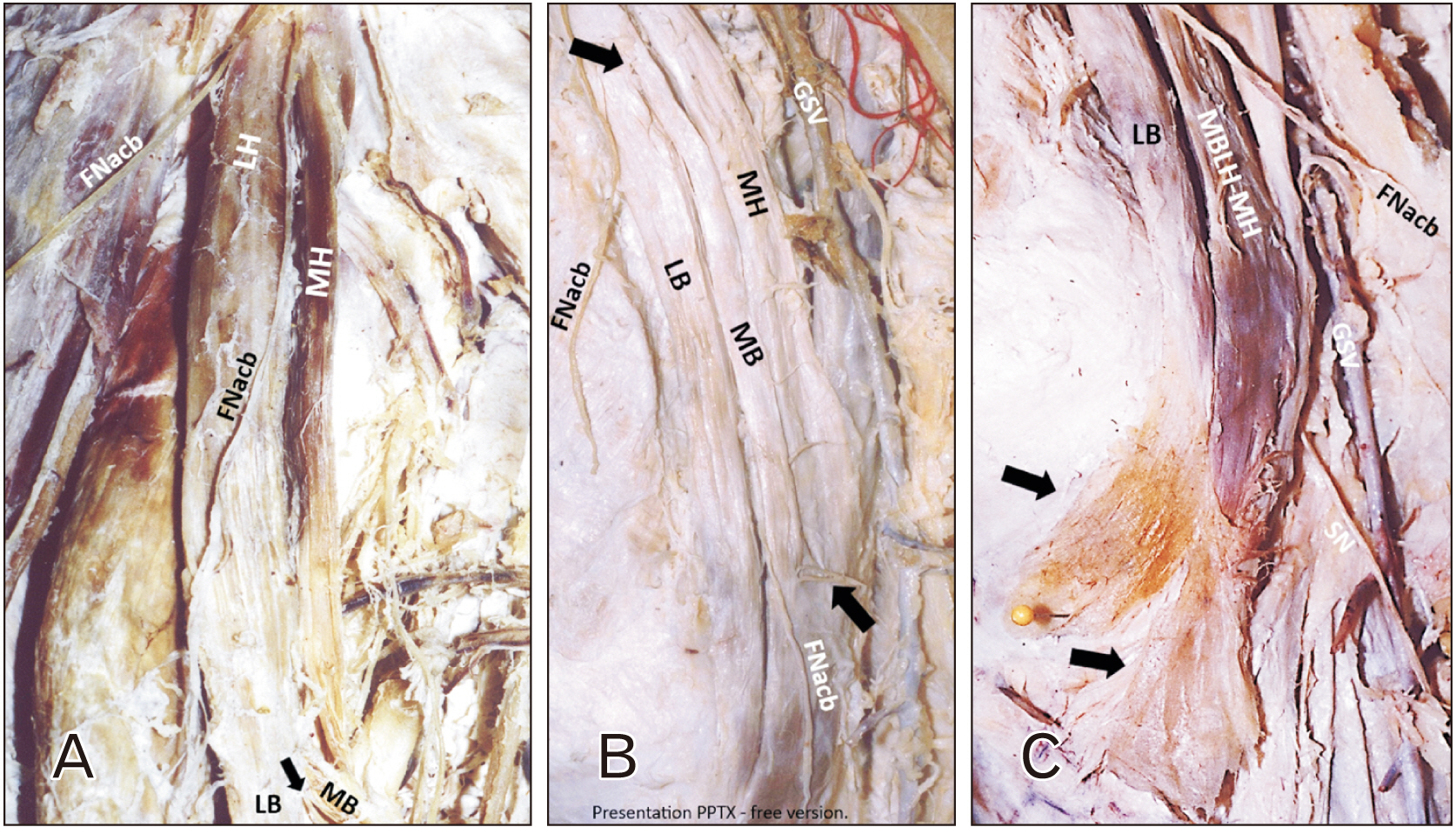
Fig. 1 (A–C) Dissection of the sartorius muscle (SM) variant and related nerves. (A) The SM division into LH and MH, and the LH division (arrow) into LB and MB. (B) The FNacb emersion (superior arrow) and the inferior black arrow depict the fusion of the MB with the MH. (C) SN-sural nerve with the GSV-great saphenous vein. Two arrows depict the superior and inferior attachments of the muscle’s components at pes anserinus. FNacb, femoral nerve anterior cutaneous branch; MH, medial head; LH, lateral head; LB, lateral bundle; MB, medial bundle; SN, saphenous nerve; GSV, great saphenous vein.
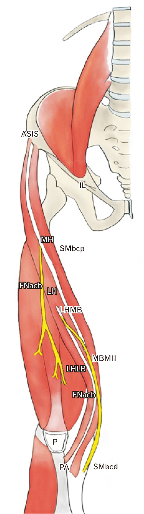
Fig. 2 Panoramic schematic view of the sartorius muscle (SM) variant. The biceps and bicaudatus SM (SMbcp and SMbcd). SM division into LH and MH, and the LH division into LB and MB. The complex MB-MH insertion into the pens anserinus (PA) inferior area. The insertion of the LB into the PA, superior area. ASIS, anterior superior iliac spine; IL, inguinal ligament; MH, medial head; LH, lateral head; MB, medial bundle; LB, lateral bundle; FNacb, the femoral nerve anterior cutaneous branch; P, patella; FN, femoral nerve.
Reference
-
References
1. Bergman RA, Afifi AK, Miyauchi R. Illustrated encyclopedia of human anatomic variation. Anatomy Atlas;2017.2. Kim J, Lee JH. 2019; A unique case of an accessory sartorius muscle. Surg Radiol Anat. 41:323–5. DOI: 10.1007/s00276-018-2155-5. PMID: 30539207.
Article3. Buckland A, Pan WR, Dhar S, Edwards G, Rozen WM, Ashton MW, Taylor GI. 2009; Neurovascular anatomy of sartorius muscle flaps: implications for local transposition and facial reanimation. Plast Reconstr Surg. 123:44–54. DOI: 10.1097/PRS.0b013e3181904bc6. PMID: 19116533.
Article4. Lin SE, Auyong DB, Dahl AB, Hanson NA. 2017; Successful continuous adductor canal block placement in a patient with absent sartorius muscle: a case report. A A Case Rep. 9:101–4. DOI: 10.1213/XAA.0000000000000538. PMID: 28410261.5. Zielinska N, Tubbs RS, Balcerzak A, Olewnik Ł. A very rare case report: accessory head of the sartorius muscle. Folia Morphol (Warsz). 2023; Feb. 22. [Epub]. https://doi.org/10.5603/FM.a2023.0014. DOI: 10.5603/FM.a2023.0014. PMID: 36811136.
Article6. Patil J, Kumar N, Swamy RS, Guru A, Mohandas Rao KG, Aithal AP. 2015; Unilateral accessory Sartorius muscle: a case report on its functional and clinical implications. Saudi J Sports Med. 15:285–7. DOI: 10.4103/1319-6308.164317.
Article7. Garbelotti JS, Rodrigues CFS, Nobeschi L, Seiji F, Olave E. 1999; Anatomical variation of the sartorius muscle. Rev Chil Anat. 17:95–7. DOI: 10.4067/S0716-98681999000100014.8. Ledouble AF. Traité des variations du système musculaire de l'homme. Tome 2;Paris: 1897.9. Macalister A. 1875; Additional observations on muscular anomalies in human anatomy. Trans R Ir Acad. 23:1–134.10. Brock GS. 1879; A two-headed sartorius. J Anat Physiol. 13(Pt 4):578. PMID: 17231291. PMCID: PMC1309846.11. Solomons JNT, Sagir A, Yazdi C. 2022; Meralgia paresthetica. Curr Pain Headache Rep. 26:525–31. DOI: 10.1007/s11916-022-01053-7. PMID: 35622311.
Article12. Chang KV, Mezian K, Naňka O, Wu WT, Lou YM, Wang JC, Martinoli C, Özçakar L. 2018; Ultrasound imaging for the cutaneous nerves of the extremities and relevant entrapment syndromes: from anatomy to clinical implications. J Clin Med. 7:457. DOI: 10.3390/jcm7110457. PMID: 30469370. PMCID: PMC6262579.
Article13. Petis S, Howard JL, Lanting BL, Vasarhelyi EM. 2015; Surgical approach in primary total hip arthroplasty: anatomy, technique and clinical outcomes. Can J Surg. 58:128–39. DOI: 10.1503/cjs.007214. PMID: 25799249. PMCID: PMC4373995.
Article14. Charalambous CP, Kwaees TA. 2013; Anatomical considerations in hamstring tendon harvesting for anterior cruciate ligament reconstruction. Muscles Ligaments Tendons J. 2:253–7. PMID: 23738306. PMCID: PMC3666537.15. Manjunath KN, Venkatesh MS, Shivaprasad A. 2018; Distal major pedicle of sartorius muscle flap: anatomical study and its clinical implications. Indian J Plast Surg. 51:40–5. DOI: 10.4103/ijps.IJPS_127_17. PMID: 29928078. PMCID: PMC5992938.
Article
- Full Text Links
-
- Actions
-
Cited
- CITED
-
- Close
- Share
-
- Similar articles
-
- Accessory Tendon of Biceps Brachii Originated from Pectoralis Major
- The Third Head of the Biceps Brachii Muscle Originated from the Pectoralis Major Muscle
- Implication of Sternalis Muscle on Staged Breast Reconstruction with Implant
- A Familial Case with Phenotypic Differences in a CAV3 Pathogenic Variant
- Clinical importance of tensor fasciae suralis arising from linea aspera along with short head of biceps femoris: a rare anomaly
