Effectiveness of digital subtraction radiography in detecting artificially created osteophytes and erosions in the temporomandibular joint
- Affiliations
-
- 1Department of Comprehensive Dentistry, The University of Texas Health Science Center, San Antonio, TX, USA. demirturk@uthscsa.edu
- 2Department of Oral and Maxillofacial Radiology, Faculty of Dentistry, Ondokuz Mayıs University, Samsun, Turkey.
- KMID: 2389890
- DOI: http://doi.org/10.5624/isd.2017.47.2.99
Abstract
- PURPOSE
Erosions and osteophytes are radiographic characteristics that are found in different stages of temporomandibular joint (TMJ) osteoarthritis. This study assessed the effectiveness of digital subtraction radiography (DSR) in diagnosing simulated osteophytes and erosions in the TMJ.
MATERIALS AND METHODS
Five intact, dry human skulls were used to assess the effectiveness of DSR in detecting osteophytes. Four cortical bone chips of varying thicknesses (0.5 mm, 1.0 mm, 1.5 mm, and 2.0 mm) were placed at the medial, central, and lateral aspects of the condyle anterior surface. Two defects of varying depth (1.0 mm and 1.5 mm) were created on the lateral, central, and medial poles of the condyles of 2 skulls to simulate erosions. Panoramic images of the condyles were acquired before and after artificially creating the changes. Digital subtraction was performed with Emago dental image archiving software. Five observers familiar with the interpretation of TMJ radiographs evaluated the images. Receiver operating characteristic (ROC) analysis was used to evaluate the diagnostic accuracy of the imaging methods.
RESULTS
The area under the ROC curve (Az) value for the overall diagnostic accuracy of DSR in detecting osteophytic changes was 0.931. The Az value for the overall diagnostic accuracy of panoramic imaging was 0.695. The accuracy of DSR in detecting erosive changes was 0.854 and 0.696 for panoramic imaging. DSR was remarkably more accurate than panoramic imaging in detecting simulated osteophytic and erosive changes.
CONCLUSION
The accuracy of panoramic imaging in detecting degenerative changes was significantly lower than the accuracy of DSR (P<.05). DSR improved the accuracy of detection using panoramic images.
Keyword
MeSH Terms
Figure
-
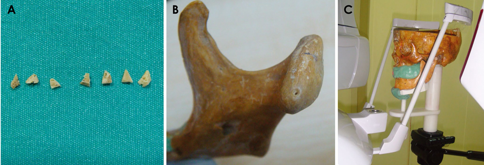
Fig. 1 A. Bone chips were used to simulate osteophyte formation on the condylar heads. B. An artificial erosion was created on the medial pole of the right condyle. C. The skull was positioned for panoramic image acquisition.
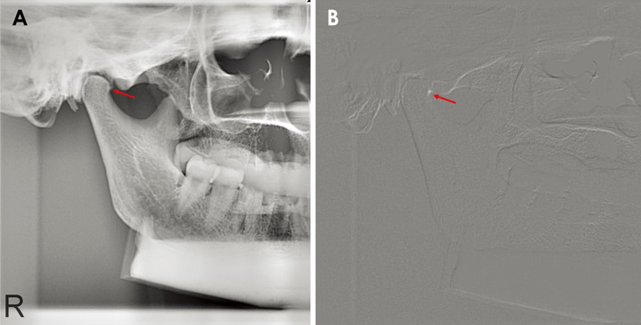
Fig. 2 A. A panoramic image shows the 2.0-mm bone chip placed on the anterolateral aspect of the right condylar head. B. A subtracted image shows the same condyle with the same bone chip.
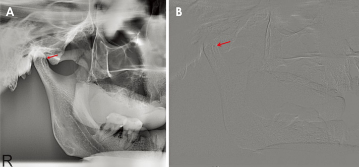
Fig. 3 A. A panoramic image shows a 1.5-mm erosion created on the medial pole of the right mandibular condyle. B. A subtracted image shows the same condyle with the erosion.
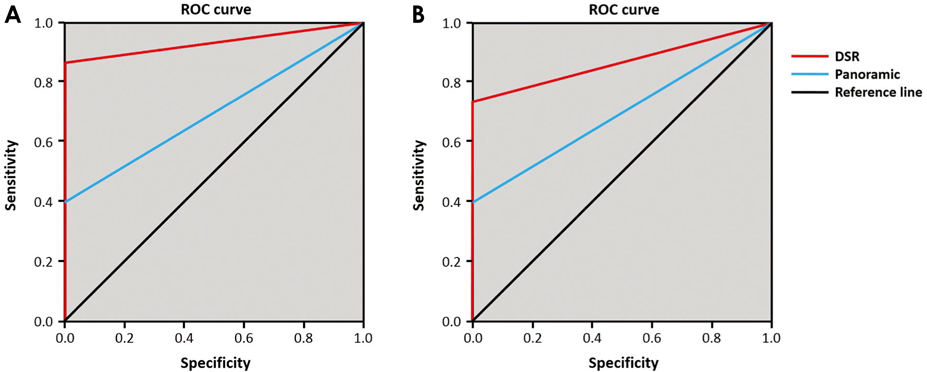
Fig. 4 A. Receiver operating characteristic (ROC) analysis of the detection of overall condylar osteophytes. Curves were plotted from the data obtained for each modality when all osteophyte sizes and locations were considered. B. ROC analysis of the detection of overall condylar erosions. Curves were plotted from the data obtained for each modality when all erosion sizes and locations were considered.
Cited by 1 articles
-
Digital subtraction radiography in TMJ imaging: A critique
Galal Omami
Imaging Sci Dent. 2017;47(3):215-217. doi: 10.5624/isd.2017.47.3.215.
Reference
-
1. Librizzi ZT, Tadinada AS, Valiyaparambil JV, Lurie AG, Mallya SM. Cone-beam computed tomography to detect erosions of the temporomandibular joint: effect of field of view and voxel size on diagnostic efficacy and effective dose. Am J Orthod Dentofacial Orthop. 2011; 140:e25–e30.
Article2. Paesani D, Westesson PL, Hatala MP, Tallents RH, Brooks SL. Accuracy of clinical diagnosis for TMJ internal derangement and arthrosis. Oral Surg Oral Med Oral Pathol. 1992; 73:360–363.
Article3. Ahmad M, Hollender L, Anderson Q, Kartha K, Ohrbach R, Truelove EL, et al. Research diagnostic criteria for temporomandibular disorders (RDC/TMD): development of image analysis criteria and examiner reliability for image analysis. Oral Surg Oral Med Oral Pathol Oral Radiol Endod. 2009; 107:844–860.
Article4. Larheim TA, Kolbenstvedt A. High-resolution computed tomography of the osseous temporomandibular joint. Some normal and abnormal appearances. Acta Radiol Diagn (Stockh). 1984; 25:465–469.5. Hussain AM, Packota G, Major PW, Flores-Mir C. Role of different imaging modalities in assessment of temporomandibular joint erosions and osteophytes: a systematic review. Dentomaxillofac Radiol. 2008; 37:63–71.
Article6. Masood F, Katz JO, Hardman PK, Glaros AG, Spencer P. Comparison of panoramic radiography and panoramic digital subtraction radiography in the detection of simulated osteophytic lesions of the mandibular condyle. Oral Surg Oral Med Oral Pathol Oral Radiol Endod. 2002; 93:626–631.
Article7. Revesz G, Kundel HL, Graber MA. The influence of structured noise on the detection of radiologic abnormalities. Invest Radiol. 1974; 9:479–486.
Article8. Brooks SL, Brand JW, Gibbs SJ, Hollender L, Lurie AG, Omnell KA, et al. Imaging of the temporomandibular joint: a position paper of the American Academy of Oral and Maxillofacial Radiology. Oral Surg Oral Med Oral Pathol Oral Radiol Endod. 1997; 83:609–618.9. Dreyer WP. Technological advances in the clinical diagnosis of periodontal diseases. Int Dent J. 1993; 43:557–566.10. Woo BM, Zee KY, Chan FH, Corbet EF. In vitro calibration and validation of a digital subtraction radiography system using scanned images. J Clin Periodontol. 2003; 30:114–118.
Article11. Hekmatian E, Sharif S, Khodaian N. Literature review digital subtraction radiography in dentistry. Dent Res J (Isfahan). 2005; 2:1–8.12. Honey OB, Scarfe WC, Hilgers MJ, Klueber K, Silveira AM, Haskell BS, et al. Accuracy of cone-beam computed tomography imaging of the temporomandibular joint: comparisons with panoramic radiology and linear tomography. Am J Orthod Dentofacial Orthop. 2007; 132:429–438.
Article13. Ludlow J, Gilbert DB, Tyndall DA, Bailey L. Analysis of condylar position change on digitally subtracted Orthophos P-4 and Sectograph zonogram images. Int J Adult Orthodon Orthognath Surg. 1995; 10:201–209.14. Larheim TA, Abrahamsson AK, Kristensen M, Arvidsson LZ. Temporomandibular joint diagnostics using CBCT. Dentomaxillofac Radiol. 2015; 44:20140235.
Article15. Mozzo P, Procacci C, Tacconi A, Martini PT, Andreis IA. A new volumetric CT machine for dental imaging based on the cone-beam technique: preliminary results. Eur Radiol. 1998; 8:1558–1564.
Article16. Arai Y, Tammisalo E, Iwai K, Hashimoto K, Shinoda K. Development of a compact computed tomographic apparatus for dental use. Dentomaxillofac Radiol. 1999; 28:245–248.
Article17. Honda K, Larheim TA, Johannessen S, Arai Y, Shinoda K, Westesson PL. Ortho cubic super-high resolution computed tomography: a new radiographic technique with application to the temporomandibular joint. Oral Surg Oral Med Oral Pathol Oral Radiol Endod. 2001; 91:239–243.
Article18. Tsiklakis K, Syriopoulos K, Stamatakis HC. Radiographic examination of the temporomandibular joint using cone beam computed tomography. Dentomaxillofac Radiol. 2004; 33:196–201.
Article19. Honda K, Larheim TA, Maruhashi K, Matsumoto K, Iwai K. Osseous abnormalities of the mandibular condyle: diagnostic reliability of cone beam computed tomography compared with helical computed tomography based on an autopsy material. Dentomaxillofac Radiol. 2006; 35:152–157.
Article20. Hintze H, Wiese M, Wenzel A. Cone beam CT and conventional tomography for the detection of morphological temporomandibular joint changes. Dentomaxillofac Radiol. 2007; 36:192–197.
Article21. Alkhader M, Ohbayashi N, Tetsumura A, Nakamura S, Okochi K, Momin MA, et al. Diagnostic performance of magnetic resonance imaging for detecting osseous abnormalities of the temporomandibular joint and its correlation with cone beam computed tomography. Dentomaxillofac Radiol. 2010; 39:270–276.
Article22. Katakami K, Shimoda S, Kobayashi K, Kawasaki K. Histological investigation of osseous changes of mandibular condyles with backscattered electron images. Dentomaxillofac Radiol. 2008; 37:330–339.
Article23. Marques AP, Perrella A, Arita ES, Pereira MF, Cavalcanti Mde G. Assessment of simulated mandibular condyle bone lesions by cone beam computed tomography. Braz Oral Res. 2010; 24:467–474.
Article24. Patel A, Tee BC, Fields H, Jones E, Chaudhry J, Sun Z. Evaluation of cone-beam computed tomography in the diagnosis of simulated small osseous defects in the mandibular condyle. Am J Orthod Dentofacial Orthop. 2014; 145:143–156.
Article
- Full Text Links
-
- Actions
-
Cited
- CITED
-
- Close
- Share
-
- Similar articles
-
- Digital subtraction radiography in TMJ imaging: A critique
- Digital contrast subtraction radiography for proximal caries diagnosis
- Cone-beam computed tomography versus digital periapical radiography in the detection of artificially created periapical lesions: A pilot study of the diagnostic accuracy of endodontists using both techniques
- Absorbed doses in organs of the head and neck from conventional temporomandibular joint tomography
- Assessment of apical root resorption using digital subtraction radiography
