Intramuscular Hematoma Following Radial Extracorporeal Shockwave Therapy for Chronic Neurogenic Heterotopic Ossification: A Case Report
- Affiliations
-
- 1Department of Rehabilitation Medicine, Kwangju Christian Hospital, Gwangju, Korea. eykang74@hanmail.net
- KMID: 2389468
- DOI: http://doi.org/10.5535/arm.2017.41.3.498
Abstract
- Extracorporeal shockwave therapy (ESWT) has been reported to be a safe and effective method for decreasing pain and relieving range of motion (ROM) limitations caused by neurogenic heterotopic ossification (NHO), though there has been no report that it might cause hematoma if applied to NHO. We hereby report a case of massive hematoma after ESWT, specifically the radial shockwave therapy (RSWT) device at both hips in a 49-year-old female patient with NHO. She had developed NHO after extensive subarachnoid hemorrhage. We had applied RSWT according to the previous report. The pain and the ROM limitations were gradually improved. Six weeks later, she reported pain and ROM limitations on the right hip. From a medial aspect, swelling and bruising of the right thigh could be seen. Magnetic resonance imaging and ultrasonography suggested a large hematoma between right hip adductor muscles. The symptoms disappeared after conservative treatment for one month, and subsequent follow-up imaging studies demonstrated resolution of the hematoma.
MeSH Terms
Figure
-
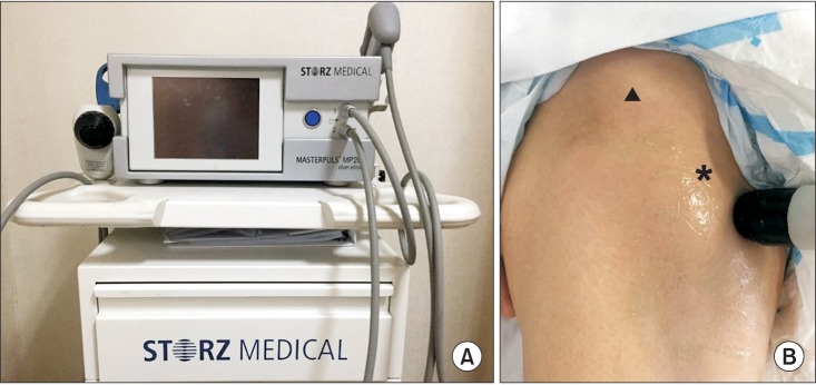
Fig. 1 (A) MP200 (Storz Medical AG, Tagerwilen, Switzerland). (B) Radial shockwave therapy was applied around a lesser trochanter (asterisk) below anterior inferior iliac spine (arrowhead).
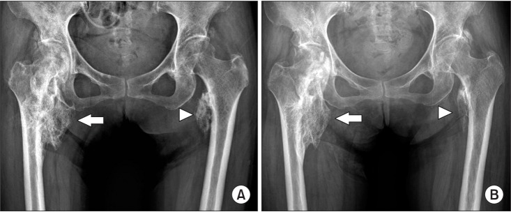
Fig. 2 Comparison of X-rays pretreatment (A) and post-treatment (B), which radial shockwave therapy revealed that the size had become slightly smaller in posttreatment on both right (arrow) and left neurogenic heterotopic ossification (arrowhead).
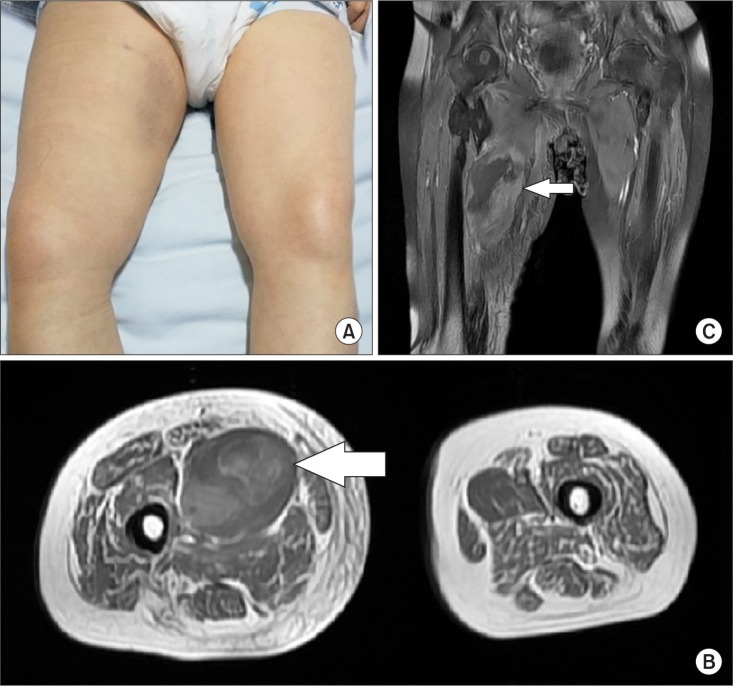
Fig. 3 (A) From a medial aspect of the right thigh, swelling and bruising were seen. Axial (B) and coronal (C) T1-weighted magnetic resonance images show an area of heterogeneous high signal intensity in the right pectineus muscle, consistent with hematoma (arrow).
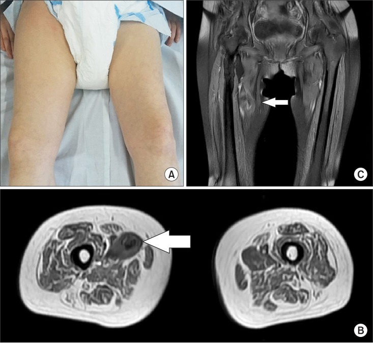
Fig. 4 (A) Swelling and bruising almost disappeared after one month of conservative treatment. Axial (B) and coronal (C) T1-weighted magnetic resonance images show that an area of hematoma had markedly decreased (arrow).
Reference
-
1. Chang KV, Chen SY, Chen WS, Tu YK, Chien KL. Comparative effectiveness of focused shock wave therapy of different intensity levels and radial shock wave therapy for treating plantar fasciitis: a systematic review and network meta-analysis. Arch Phys Med Rehabil. 2012; 93:1259–1268. PMID: 22421623.
Article2. Cacchio A, Paoloni M, Barile A, Don R, de Paulis F, Calvisi V, et al. Effectiveness of radial shock-wave therapy for calcific tendinitis of the shoulder: single-blind, randomized clinical study. Phys Ther. 2006; 86:672–682. PMID: 16649891.
Article3. Brissot R, Lassalle A, Vincendeau S, Polard JL, Fouche M, Ninubona D, et al. Treatment of heterotopic ossification by extracorporeal shock wave: 26 patients. Ann Readapt Med Phys. 2005; 48:581–589. PMID: 15993976.4. Reznik JE, Milanese S, Golledge J, Biros E, Gordon S, Galea MP. Extracorporeal shock wave therapy as a treatment for heterotopic ossification. Phys Ther Rev. 2013; 18:300–307.
Article5. Choi YM, Hong SH, Lee CH, Kang JH, Oh JS. Extracorporeal shock wave therapy for painful chronic neurogenic heterotopic ossification after traumatic brain injury: a case report. Ann Rehabil Med. 2015; 39:318–322. PMID: 25932431.
Article6. Haake M, Boddeker IR, Decker T, Buch M, Vogel M, Labek G, et al. Side-effects of extracorporeal shock wave therapy (ESWT) in the treatment of tennis elbow. Arch Orthop Trauma Surg. 2002; 122:222–228. PMID: 12029512.7. Sistermann R, Katthagen BD. Complications, side-effects and contraindications in the use of medium and high-energy extracorporeal shock waves in orthopedics. Z Orthop Ihre Grenzgeb. 1998; 136:175–181. PMID: 9615982.8. Csaszar NB, Angstman NB, Milz S, Sprecher CM, Kobel P, Farhat M, et al. Radial Shock Wave Devices Generate Cavitation. PLoS One. 2015; 10:e0140541. PMID: 26509573.
Article
- Full Text Links
-
- Actions
-
Cited
- CITED
-
- Close
- Share
-
- Similar articles
-
- Extracorporeal Shock Wave Therapy for Painful Chronic Neurogenic Heterotopic Ossification After Traumatic Brain Injury: A Case Report
- Heterotopic Ossification Mimics Neurogenic Tumor: A Case Report
- Osteomyelitis in Heterotopic Ossification after Trochanteric Pressure Sore Reconstruction: A Case Report
- Heterotopic Ossification of a Partially Ruptured Achilles Tendon (A Case Report)
- Extracorporeal Shock Wave Therapy for Painful Heterotopic Ossification after Traumatic Transtibial Amputation
