Effects of Exercise on Neuropathy in Streptozotocin-Induced Diabetic Rats
- Affiliations
-
- 1Department of Physical and Rehabilitation Medicine, Inha University, Incheon, Korea. rmkmo@inha.ac.kr
- KMID: 2389455
- DOI: http://doi.org/10.5535/arm.2017.41.3.402
Abstract
OBJECTIVE
To evaluate the effects of early regular exercise and to assess the electrophysiological and histopathological findings of the rat tail nerve in relation to the timing of exercise training for swimming exercise in rats with diabetic neuropathy.
METHODS
We used 70 Sprague-Dawley male rats, and the experimental group comprised 60 rats, and the control group comprised 10 rats. Diabetes was induced by intraperitoneal injection of streptozotocin. Blood glucose concentrations were measured in tail vein blood samples. The experimental group was divided into 6 subgroups according to insulin treatment and swimming exercise: group 1, diabetic control; group 2, insulin treated; group 3, insulin untreated with early swimming exercise; group 4, insulin treated and early swimming exercise; group 5, insulin treated and late swimming exercise; and group 6, insulin untreated with late swimming exercise. Sensory and motor nerve conduction studies were performed weekly up to the 13th week using rat tail nerves. The effect on structural diabetic neuropathy was assessed by morphometry and ultrastructural examination of the rat tail nerve fiber at the 14th week.
RESULTS
An exercise effect was observed in the insulin treated groups, but it was not observed in the insulin untreated groups. The sensory nerve conduction study in the rat tail revealed significantly prolonged latency and decreased amplitude in groups 1 and 6, and a further delay was observed in group 5 when compared to group 4. Decreased thickness of myelin was found in groups 1 and 6 through morphometry.
CONCLUSION
Early regular exercise programs in addition to conventional insulin treatment may retard the progression of diabetic peripheral neuropathy.
MeSH Terms
Figure
-

Fig. 1 Flow chart of the experiment.
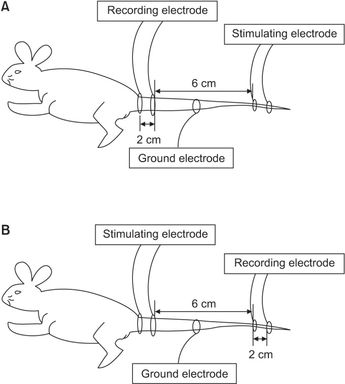
Fig. 2 (A) Measurements of the orthodromic sensory nerve conduction study. (B) Measurements of the motor nerve conduction study.
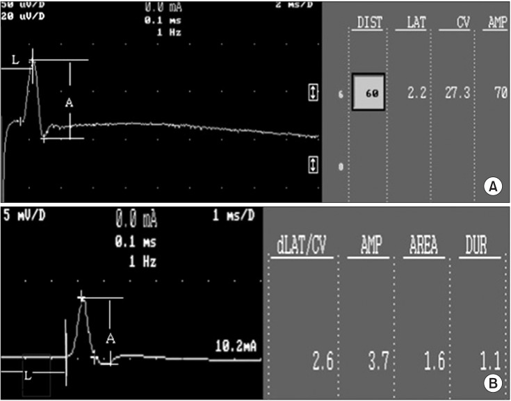
Fig. 3 (A) An example of sensory nerve action potential of the rat tail nerve. (B) An example of compound muscle action potential of the rat tail nerve. A, amplitude; L, latency.
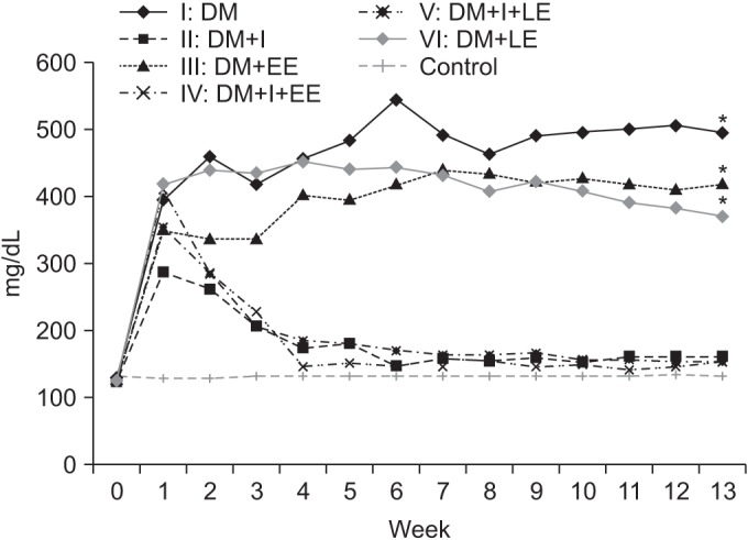
Fig. 4 Effects of insulin and exercise treatment on blood glucose levels. A significant difference was observed between insulin untreated groups and insulin treated groups from the 3rd week (*p<0.05, difference of blood glucose levels between groups 2, 4, 5 and groups 1, 3, 6). Blood glucose level was decreased significantly in the insulin treated groups in comparison with the insulin untreated group. DM, diabetes mellitus; I, insulin treated; EE, early exercise; LE, late exercise.
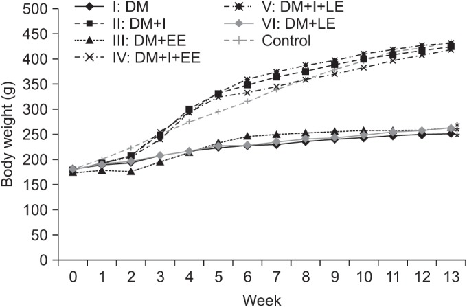
Fig. 5 Effects of insulin and exercise treatment on body weight. A significant difference was observed between the insulin untreated groups and the insulin treated groups from the 4th week (*p<0.05, difference of body weight between groups 2, 4, 5 and groups 1, 3, 6). Body weight was decreased significantly in the insulin untreated groups in comparison with the insulin treated group. DM, diabetes mellitus; I, insulin treated; EE, early exercise; LE, late exercise.
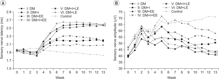
Fig. 6 (A) Effects of insulin and exercise treatment on sensory nerve latency. A significant difference was observed between the insulin untreated groups and the insulin treated groups from the 5th week (*p<0.05, difference of distal latency in sensory nerve conduction study between groups 2, 4, 5 and groups 1, 3, 6). Sensory nerve latency was decreased significantly in the insulin treated early exercise group in comparison with the other insulin treated group from the 7th week. (B) Effects of insulin and exercise treatment on sensory nerve amplitude. A significant difference was observed between the insulin treated early exercise group and the insulin treated non-exercise group from the 5th week. Sensory nerve amplitude was increased significantly in the insulin treated early exercise group in comparison with the other insulin treated late exercise group from the 7th week (*p<0.05, difference of amplitude in sensory nerve conduction study between groups 2, 4, 5 and groups 1, 3, 6). DM, diabetes mellitus; I, insulin treated; EE, early exercise; LE, late exercise.
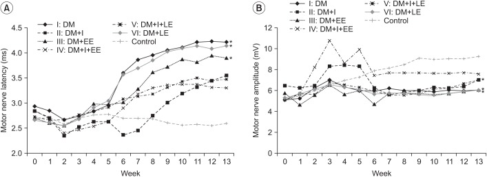
Fig. 7 (A) Effects of insulin and exercise treatment on motor nerve latency. A significant difference was observed between the insulin untreated groups and the insulin treated groups from the 5th week (*p<0.05, difference of distal latency in motor nerve conduction study between groups 2, 4, 5 and groups 1, 3, 6). No significantly different values were noted in the insulin untreated groups or the insulin treated groups. (B) Effects of insulin and exercise treatment on motor nerve amplitude. A significant difference was observed between the insulin treated with early exercise group and the other groups at the 3rd week and from the 7th to the 9th week (*p<0.05, difference of amplitude in motor nerve conduction study between groups 2, 4, 5 and groups 1, 3, 6). No significant differences were noted from the values in the insulin untreated groups. DM, diabetes mellitus; I, insulin treated; EE, early exercise; LE, late exercise.
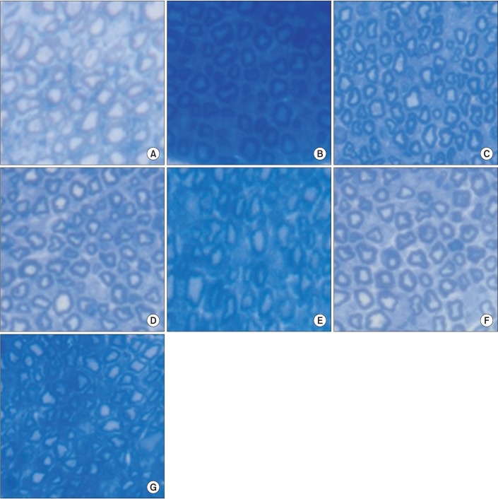
Fig. 8 Microscopic examination of rat tail nerves (toluidine blue, ×400): (A) normal control, (B) group 1, (C) group 2, (D) group 3, (E) group 4, (F) group 5, and (G) group 6. There was no evidence of demyelination or axonopathy in all groups.
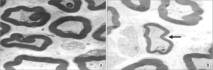
Fig. 9 Electron microscopic findings of rat tail nerves (×5,000): (A) control group and (B) group 1. A disproportionately shrunk thin myelin sheath was observed (arrow).
Reference
-
1. Pyun SB, Kwon HK, Uhm CS. Effect of exercise on reinnervating soleus muscle after sciatic nerve injury in rats. J Korean Acad Rehabil Med. 1999; 23:1063–1075.2. Dumitru D. Electrodiagnostic medicine. 1st ed. Philadelphia: Mosby;1995. p. 821–824.3. Dyck PJ, Thomas PK. Diabetic neuropathy. 2nd ed. Philadelphia: W.B. Saunders;1999. p. 222–236. p. 330–340.4. Singleton JR, Smith AG, Marcus RL. Exercise as therapy for diabetic and prediabetic neuropathy. Curr Diab Rep. 2015; 15:120. PMID: 26538074.
Article5. Malysz T, Ilha J, Nascimento PS, De Angelis K, Schaan BD, Achaval M. Beneficial effects of treadmill training in experimental diabetic nerve regeneration. Clinics (Sao Paulo). 2010; 65:1329–1337. PMID: 21340223.
Article6. van Meeteren NL, Brakkee JH, Helders PJ, Wiegant VM, Gispen WH. Functional recovery from sciatic nerve crush lesion in the rat correlates with individual differences in responses to chronic intermittent stress. J Neurosci Res. 1997; 48:524–532. PMID: 9210522.
Article7. Kwon HK, Lee HJ, Yim SK, Lee SR. Electrophysiologic assessement of axonopathy and demyelination in diabetic neuropathy according to the severity. J Korean Acad Rehabil Med. 2002; 26:50–54.8. Kim MO, Kim SJ, Choi HC, Roh GH, Kim SY. Histochemical findings of soleus in relation to the severity of injury and duration of exercise in sciatic nerve injured rats. J Korean Acad Rehabil Med. 2003; 27:727–734.9. Abu-Shakra SR, Cornblath DR, Avila OL, Chaudhry V, Freimer M, Glass JD, et al. Conduction block in diabetic neuropathy. Muscle Nerve. 1991; 14:858–862. PMID: 1922181.
Article10. Said G. Diabetic neuropathy: a review. Nat Clin Pract Neurol. 2007; 3:331–340. PMID: 17549059.11. Park DW, Nam KS, Kim SC, Park SI, Choi E, Lee YG. Significance of amplitude and area ratio of compound muscle action potential in diagnosis of diabetic neuropathy. J Korean Acad Rehabil Med. 2001; 25:615–620.12. Park BK, Kim SJ. Response of peripheral nerve to transient ischemia in streptozotocin-induced diabetic rats. J Korean Acad Rehabil Med. 1993; 17:392–405.13. Kim CH, Choi HC, Roh GH. The preventive effect of nimodipine on the cisplatin induced neuropathy. J Korean Acad Rehabil Med. 2003; 27:90–95.14. Gulsen I, Demiroglu M, Aycan A, Ucler R, Alaca I, Orhon ZN, et al. Effects of low-intensity treadmill exercise on sciatic nerve in experimental diabetic neuropathy. Anal Quant Cytopathol Histpathol. 2016; 38:95–102. PMID: 27386630.15. Kim WS, Lee SU. Harmful effect of land-based endurance exercise in rats with diabetic nerve. Med Sci Sports Exerc. 2010; 42:1625–1631. PMID: 20142779.
Article16. Kim MO, Yoon JS, Kwak JR, Choi HC, Roh GH, Kim SJ. Effect of therapeutic exercise according to degree of injury in sciatic nerve damaged rat. J Korean Acad Rehabil Med. 2001; 25:466–473.17. Dyck PJ, Davies JL, Wilson DM, Service FJ, Melton LJ 3rd, O'Brien PC. Risk factors for severity of diabetic polyneuropathy: intensive longitudinal assessment of the Rochester Diabetic Neuropathy Study cohort. Diabetes Care. 1999; 22:1479–1486. PMID: 10480512.
Article18. Eugene B, Abthony SF, Dennis LK, Stephen LH, Dan LL. Harrison's principles of internal medicine. 15th ed. McGraw-Hill;2001. p. 2109–2137.19. Hotta N, Koh N, Sakakibara F, Nakamura J, Hamara Y, Hara T, et al. Effects of propionyl-L-carnitine and insulin on the electroretinogram, nerve conduction and nerve blood flow in rats with streptozotocin-induced diabetes. Pflugers Arch. 1996; 431:564–570. PMID: 8596700.
Article
- Full Text Links
-
- Actions
-
Cited
- CITED
-
- Close
- Share
-
- Similar articles
-
- Protective Effect of Melatonin on Neuropathy in Streptozotocin-Induced Diabetic Rats
- Proliferation of Cultured Vascular Smooth Muscle Cells(VSMCs) Obtained from Aortas of Insulin Dependent Diabetic Rats
- The Effects of Nerve Growth Factor on Satellite Cell of the Dorsal Rott Ganglia in Streptozotocin-induced Diabetic Rats
- Alteration of Nerve Growth Factor in the Penis of Rats with Streptozotocin-induced Diabetes
- The Morphological Studies on the Effects of Nerve Growth Factor on the Schwann Cell in the Diabetic Neuropathy in the Rats
