Use of artificial palate for improving facial support in the fabrication of a maxillary obturator: A case report
- Affiliations
-
- 1Department of Prosthodontics, Dong-a University, Busan, Republic of Korea.
- 2Department of Oral and Maxillofacial Surgery, Implant Clinics, Dong-a University, Busan, Republic of Korea. omsbjkim@dau.ac.kr
- KMID: 2387206
- DOI: http://doi.org/10.4047/jkap.2017.55.3.319
Abstract
- Patients with maxillectomy defects predisposed to not only difficulty in deglutition, mastication, speech but also psychological depression from impaired facial esthetics that affect life quality. Obturator prostheses play a important role in restoring the lost form, function and the quality of life for patients with maxillectomy defects. This clinical report presents the simplified approach to predict the degree of adequate facial support by Artificial palate which reflected from a maxillary interim obturator during the stabilization period after maxillectomy.
MeSH Terms
Figure
-

Fig. 1 Initial photographs. (A) Frontal view, (B) Intraoral maxillary occlusal view, (C) Post-operative panoramic radiograph.
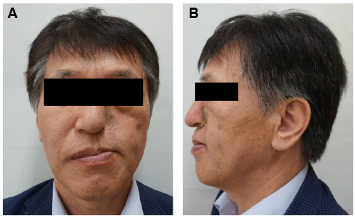
Fig. 2 Extraoral photographs. (A) Frontal view, (B) Lateral view.
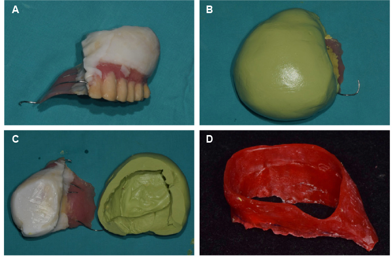
Fig. 3 The artificial palate fabrication. (A) Existing obturator prosthesis, (B) The existing obturator prosthesis placed into the silicone mold, (C) Silicone mold, (D) Artificial palate.
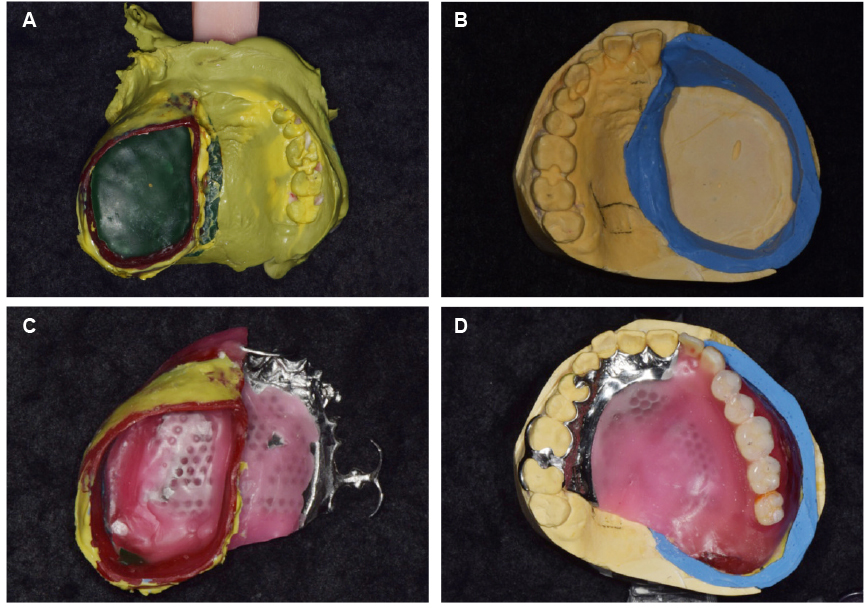
Fig. 4 (A) Artificial palate assembly made by attaching wax material to the bottom of artificial palate and pick up impression using artificial palate assembly, (B) Master cast, (C) Metal framework with artificial palate, (D) Wax denture.
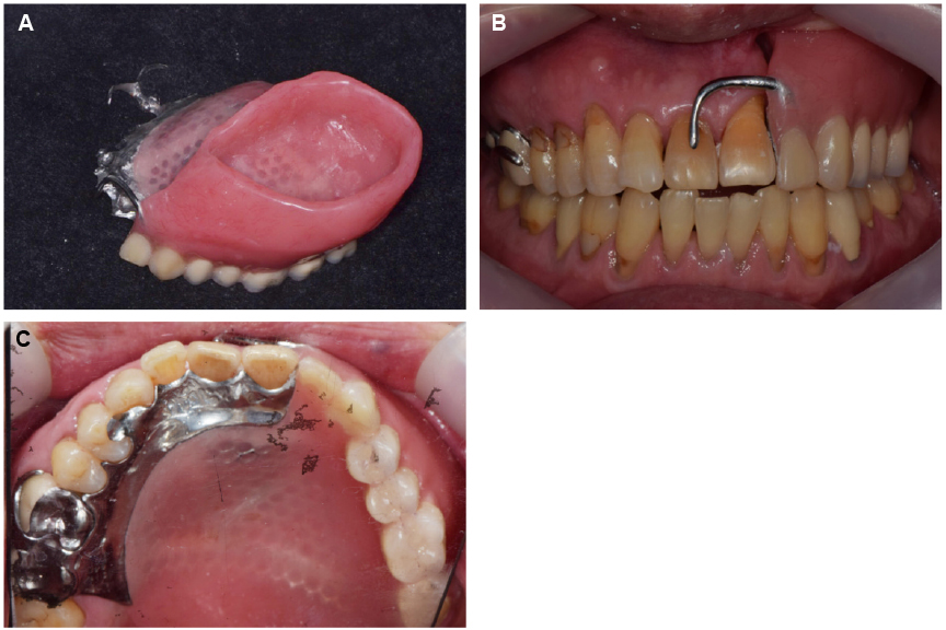
Fig. 5 5. Intraoral photographs after placement of definitive prosthesis. (A) Definitive obturator, (B) Frontal view, (C) Intraoral maxillary occlusal view.
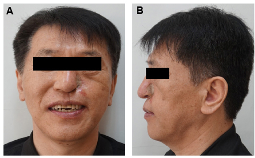
Fig. 6 Extraoral photographs after placement of definitive prosthesis. (A) Frontal view, (B) Lateral view.
Reference
-
1. Tang JA, Rieger JM, Wolfaardt JF. A review of functional outcomes related to prosthetic treatment after maxillary and mandibular reconstruction in patients with head and neck cancer. Int J Prosthodont. 2008; 21:337–354.2. Kornblith AB, Zlotolow IM, Gooen J, Huryn JM, Lerner T, Strong EW, Shah JP, Spiro RH, Holland JC. Quality of life of maxillectomy patients using an obturator prosthesis. Head Neck. 1996; 18:323–334.
Article3. Kouyoumdjian JH, Chalian VA. An interim obturator prosthesis with duplicated teeth and palate. J Prosthet Dent. 1984; 52:560–562.
Article4. DaBreo EL, Chalian VA, Lingeman R, Reisbick MH. Prosthetic and surgical management of osteogenic sarcoma of the maxilla. J Prosthet Dent. 1990; 63:316–320.
Article5. Keyf F. Obturator prostheses for hemimaxillectomy patients. J Oral Rehabil. 2001; 28:821–829.
Article6. Kaires AK. Effect of partial denture design on bilateral force distribution. J Prosthet Dent. 1956; 6:373–385.
Article7. Kwon HB, Lee JB, Yim SH. Patients' satisfaction on the obturators with different extension heights into defects after maxillectomy. J Korean Acad Prosthodont. 2010; 48:41–47.
Article8. Hanawa S, Kitaoka A, Koyama S, Sasaki K. Influence of maxillary obturator prostheses on facial morphology in patients with unilateral maxillary defects. J Prosthet Dent. 2015; 113:62–70.
Article
- Full Text Links
-
- Actions
-
Cited
- CITED
-
- Close
- Share
-
- Similar articles
-
- Fabrication of closed hollow obturator for hard palate defect patient undergone maxillectomy
- Prosthetic treatment of velopharyngeal insufficiency using maxillary obturator in an edentulous patient with Passavant’s ridge
- Fabrication of implant-associated obturator after extraction of abutment teeth: a case report
- Prosthetic rehabilitation of partially edentulous patient after maxillectomy: A case report
- Prosthetic rehabilitation by double-processing technique for edentulous patient with soft palate defect after maxillectomy: A case report
