Association of Carotid Artery Parameters of Atherosclerosis in Coronary Artery Disease
- Affiliations
-
- 1Division of Cardiology, Department of Internal Medicine, Kosin University School of Medicine, Busan, Korea. kyoungim74@gmail.com
Abstract
- BACKGROUND
Although carotid intima-media thickness (IMT) is the most commonly used ultrasonic measurement of atherosclerosis, plaque burden can be also assessed by ultrasound (US). We investigated the relationship between IMT, total plaque area (TPA) and total plaque volume (TPV) in patients with coronary artery disease (CAD).
METHODS
One hundred and seven patients with suspected CAD and carotid plaques identified by duplex ultrasound underwent 3-dimensional US and coronary angiography. The mean IMT, TPA, and TPV were analyzed for patients with CAD according to the severity of CAD.
RESULTS
In the 107 participants, IMT, TPA and TPV averaged 0.90 +/- 0.26 mm, 0.42 +/- 0.39 cm2 and 237.0 +/- 301.2 mm3, respectively. We found significant correlations for mean IMT : TPA, mean IMT : TPV and TPA : TPV of 0.448, 0.587 and 0.873, respectively (all p < 0.005). Although there was no significant association of IMT and the severity of CAD, TPA and TPV showed significant positive correlation with CAD severity (r = 0.340, p = 0.0003 for TPA and r = 0.465, p < 0.0001 for TPV). Multivariate linear regression analysis showed age was the only significant attributor to IMT, TPA, and TPV. Mean IMT was significantly associated only with hypertension. TPA was significantly associated with male sex, hypertension, and low density lipoprotein-cholesterol (LDL-C). TPV was significantly associated with male sex, C-reactive protein, and LDL-C.
CONCLUSION
Although there were significant correlations among the various US measures of carotid artery morphology, there seemed to be different biological determinants of IMT, TPA, and TPV. We might need to be selective about the particular measurements for specific applications.
Keyword
MeSH Terms
Figure
-
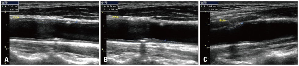
Fig. 1 The process of measurement of total plaque area (TPA) by manual planimetry. This patient showed diffuse plaques in near (A) and far (B) wall of the common carotid artery and carotid bulb (C). Measurement of plaque area was acquired from tracing the each plaque border, and TPA was the sum of the areas of all plaques (A + B + C).
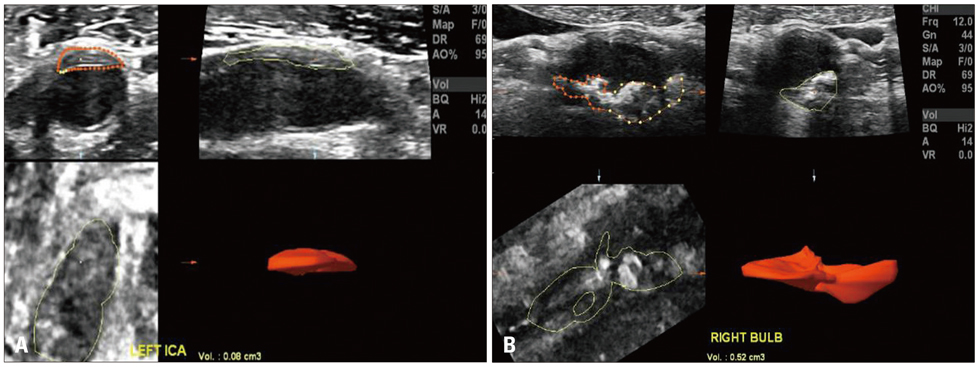
Fig. 2 The process of measurement of total plaque volume (TPV) by manual planimetry. Three-dimensional ultrasound images of plaque volumes were acquired from the longitudinal and cross sectional carotid scans. Plaque boundaries were traced using a mouse driven cross-haired cursor and cross-sectional area of the plaque was delineated from 8-12 transverse sections of the image between the edges of the plaque, and the mean plaque volume value of 3 measurements of each plaque was used for analysis. In this patient, plaques were visualized in left internal carotid artery (A) and right bulb (B), and TPV was the sum of the areas of all plaques (A + B).
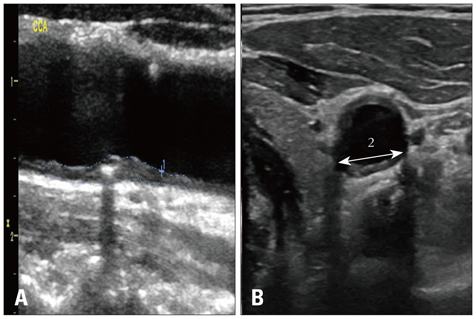
Fig. 3 Calculation of a modified plaque volume as the 2 dimensional ultrasound plaque area in the longitudinal plane (A) multiplied by plaque width (B) measured in the cross sectional plane (1 × 2).
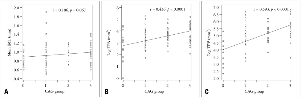
Fig. 4 Linear regression analysis among the mean carotid intima-media thickness (IMT) (A), total plaque area (TPA) (B), total plaque volume (TPV) (C), and the severity of the coronary artery disease (CAD). Although there was no significant association of IMT and the severity of CAD (group 0 having normal coronary arteries, group 1 with one vessel CAD, group 2 with two vessel CAD, and group 3 with three vessel CAD), TPA and TPV showed significant positive correlation with CAD severity (r = 0.340, p = 0.0003 for TPA and r = 0.465, p < 0.0001 for TPV). CAG: coronary angiography.

Fig. 5 Bland-Altman's plot of the difference between plaque volume (PV) measured by 3-dimensional (3D) and 2-dimensional (2D) method. A solid horizontal line gives the mean difference of 4.8 mm3 and the dotted horizontal lines give the 95% limit of agreement from -171.8 to 181.5 mm3. The difference is calculated by subtracting 2D PV from the 3D PV. The 95% limit of agreement is drawn at mean difference plus and minus 1.96 times the standard deviation (SD) of the differences.
Reference
-
1. O'Leary DH, Polak JF, Kronmal RA, Manolio TA, Burke GL, Wolfson SK Jr. Cardiovascular Health Study Collaborative Research Group. Carotid-artery intima and media thickness as a risk factor for myocardial infarction and stroke in older adults. N Engl J Med. 1999; 340:14–22.2. Chambless LE, Heiss G, Folsom AR, Rosamond W, Szklo M, Sharrett AR, Clegg LX. Association of coronary heart disease incidence with carotid arterial wall thickness and major risk factors: the Atherosclerosis Risk in Communities (ARIC) Study, 1987-1993. Am J Epidemiol. 1997; 146:483–494.
Article3. Yoon HJ, Jeong MH, Kim KH, Ahn Y, Cho JG, Park JC, Kang JC, Bae JH. Carotid intima-media thickness, not carotid plaque, is associated with large territory cerebral infarction in patients with ischemic stroke. Korean Circ J. 2010; 40:272–276.
Article4. Spence JD, Eliasziw M, DiCicco M, Hackam DG, Galil R, Lohmann T. Carotid plaque area: a tool for targeting and evaluating vascular preventive therapy. Stroke. 2002; 33:2916–2922.5. Inaba Y, Chen JA, Bergmann SR. Carotid plaque, compared with carotid intima-media thickness, more accurately predicts coronary artery disease events: a meta-analysis. Atherosclerosis. 2012; 220:128–133.
Article6. Wendelhag I, Wiklund O, Wikstrand J. On quantifying plaque size and intima-media thickness in carotid and femoral arteries. Comments on results from a prospective ultrasound study in patients with familial hypercholesterolemia. Arterioscler Thromb Vasc Biol. 1996; 16:843–850.
Article7. Spence JD. Technology Insight: ultrasound measurement of carotid plaque--patient management, genetic research, and therapy evaluation. Nat Clin Pract Neurol. 2006; 2:611–619.
Article8. Spence JD, Malinow MR, Barnett PA, Marian AJ, Freeman D, Hegele RA. Plasma homocyst(e)ine concentration, but not MTHFR genotype, is associated with variation in carotid plaque area. Stroke. 1999; 30:969–973.
Article9. Hegele RA, Ban MR, Anderson CM, Spence JD. Infection-susceptibility alleles of mannose-binding lectin are associated with increased carotid plaque area. J Investig Med. 2000; 48:198–202.10. Spence JD, Ban MR, Hegele RA. Lipoprotein lipase (LPL) gene variation and progression of carotid artery plaque. Stroke. 2003; 34:1176–1180.
Article11. Steinke W, Hennerici M. Three-dimensional ultrasound imaging of carotid artery plaques. J Cardiovasc Technol. 1989; 8:15–22.12. Delcker A, Diener HC. Quantification of atherosclerotic plaques in carotid arteries by three-dimensional ultrasound. Br J Radiol. 1994; 67:672–678.
Article13. Palombo C, Kozakova M, Morizzo C, Andreuccetti F, Tondini A, Palchetti P, Mirra G, Parenti G, Pandian NG. Ultrafast three-dimensional ultrasound: application to carotid artery imaging. Stroke. 1998; 29:1631–1637.14. Schminke U, Hilker L, Motsch L, Griewing B, Kessler C. Volumetric assessment of plaque progression with 3-dimensional ultrasonography under statin therapy. J Neuroimaging. 2002; 12:245–251.
Article15. Landry A, Fenster A. Theoretical and experimental quantification of carotid plaque volume measurements made by three-dimensional ultrasound using test phantoms. Med Phys. 2002; 29:2319–2327.
Article16. Landry A, Spence JD, Fenster A. Measurement of carotid plaque volume by 3-dimensional ultrasound. Stroke. 2004; 35:864–869.
Article17. Vermeersch SJ, Rietzschel ER, De Buyzere ML, Van Bortel LM, D'Asseler Y, Gillebert TC, Verdonck PR, Segers P. Validation of a new automated IMT measurement algorithm. J Hum Hypertens. 2007; 21:976–978.
Article18. Touboul PJ, Hennerici MG, Meairs S, Adams H, Amarenco P, Bornstein N, Csiba L, Desvarieux M, Ebrahim S, Hernandez Hernandez R, Jaff M, Kownator S, Naqvi T, Prati P, Rundek T, Sitzer M, Schminke U, Tardif JC, Taylor A, Vicaut E, Woo KS. Mannheim carotid intima-media thickness and plaque consensus (2004-2006-2011). An update on behalf of the advisory board of the 3rd, 4th and 5th watching the risk symposia, at the 13th, 15th and 20th European Stroke Conferences, Mannheim, Germany, 2004, Brussels, Belgium, 2006, and Hamburg, Germany, 2011. Cerebrovasc Dis. 2012; 34:290–296.
Article19. Egger M, Spence JD, Fenster A, Parraga G. Validation of 3D ultrasound vessel wall volume: an imaging phenotype of carotid atherosclerosis. Ultrasound Med Biol. 2007; 33:905–914.
Article20. Ludwig M, Zielinski T, Schremmer D, Stumpe KO. Reproducibility of 3-dimensional ultrasound readings of volume of carotid atherosclerotic plaque. Cardiovasc Ultrasound. 2008; 6:42.
Article21. Spence JD, Hegele RA. Noninvasive phenotypes of atherosclerosis: similar windows but different views. Stroke. 2004; 35:649–653.22. Finn AV, Kolodgie FD, Virmani R. Correlation between carotid intimal/medial thickness and atherosclerosis: a point of view from pathology. Arterioscler Thromb Vasc Biol. 2010; 30:177–181.
Article23. Brook RD, Bard RL, Patel S, Rubenfire M, Clarke NS, Kazerooni EA, Wakefield TW, Henke PK, Eagle KA. A negative carotid plaque area test is superior to other noninvasive atherosclerosis studies for reducing the likelihood of having underlying significant coronary artery disease. Arterioscler Thromb Vasc Biol. 2006; 26:656–662.
Article24. O'Leary DH, Bots ML. Imaging of atherosclerosis: carotid intima-media thickness. Eur Heart J. 2010; 31:1682–1689.25. Lorenz MW, Markus HS, Bots ML, Rosvall M, Sitzer M. Prediction of clinical cardiovascular events with carotid intima-media thickness: a systematic review and meta-analysis. Circulation. 2007; 115:459–467.
Article
- Full Text Links
-
- Actions
-
Cited
- CITED
-
- Close
- Share
-
- Similar articles
-
- Carotid ultrasound in patients with coronary artery disease
- Relationship Between Carotid and Coronary Atherosclerosis in the Elderly
- Neutrophil-to-Lymphocyte Ratio for Risk Assessment in Coronary Artery Disease and Carotid Artery Atherosclerosis
- Correlation between Intima-Media Thickness in Carotid Artery and the Extent of Coronary Atherosclerosis
- Noninvasive surrogates in assessing the severity of coronary atherosclerosis
