Seeding Metastasis of Chromophobe Renal Cell Carcinoma after Robot-Assisted Laparoscopic Partial Nephrectomy
- Affiliations
-
- 1Department of Radiology, Seoul St. Mary's Hospital, College of Medicine, The Catholic University of Korea, Seoul, Korea. cmh@catholic.ac.kr
- KMID: 2385611
- DOI: http://doi.org/10.13104/imri.2017.21.2.119
Abstract
- Chromophobe renal cell carcinoma (RCC) is an uncommon subtype of RCC having a better prognosis than clear cell RCC. Although there are several reports of seeding metastasis of RCC after biopsy, seeding metastasis of chromophobe RCC after surgical resection has seldom been reported. Here, we describe a case of multiple seeding metastases in the abdomen and pelvis 78 months after robot-assisted laparoscopic partial nephrectomy, without prior history of biopsy for chromophobe RCC in the right kidney. As magnetic resonance imaging (MRI) of the pelvic mass showed a similar appearance to the primary renal mass and displayed separate margins with the rectum and prostate gland, we were able to make a diagnosis before pathologic confirmation.
Keyword
MeSH Terms
Figure
-

Fig. 1 A 71-year-old man with an 11.4-cm-sized mass in the pelvic cavity. (a) Pelvic CT scan showed a mass with lobulated contour with heterogeneous enhancement and a non-enhancing portion in the center. The mass compressed the urinary bladder and the rectum. (b) Abdominal CT scan showed another similar mass (arrow) in the right upper quadrant (RUQ) of the abdomen.
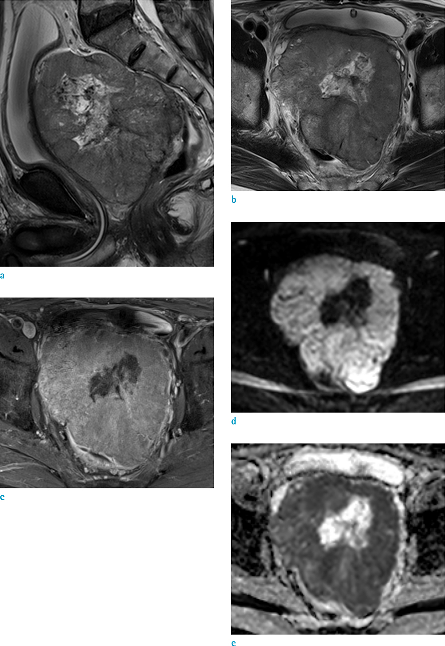
Fig. 2 A 71-year-old man with an 11.4-cm-sized mass in the pelvic cavity. (a) T2-wegithed sagittal image showed that a huge mass compressed the prostate gland, whereas the rectum had a clear margin. (b) T2-weighted axial image showed low signal intensity in peripheral portion and high signal intensity in central portion of the mass, with thin low signal intensity capsule. (c) Contrast-enhanced T1-weighted axial image displayed heterogeneous enhancement of the mass. Diffusion-weighted image (d) and apparent diffusion coefficient (ADC) map (e) showed definite diffusion restriction in peripheral solid portion.

Fig. 3 Initial MRI of right kidney mass that was confirmed as chromophobe renal cell carcinoma. The mass showed lower signal intensity than the renal parenchyma, with a central high signal intensity portion and thin low signal intensity capsule, on T2-weighted coronal image (a). Contrast-enhanced T1-weighted coronal image (b) showed the spoke-wheel-like enhancement in the mass (arrow).
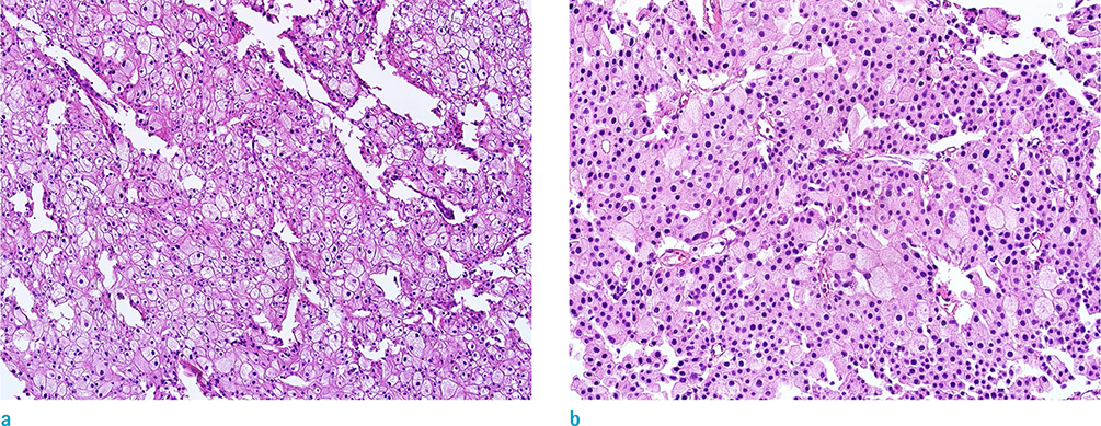
Fig. 4 The specimens obtained by partial resection of right kidney (a) and ultrasound-guided transrectal biopsy (b). The tumor showed typical features of chromophobe RCC, including pale eosinophilic cytoplasm with distinct cell membranes and perinuclear halos (Hematoxylin & Eosin staining, × 400).
Reference
-
1. Cheville JC, Lohse CM, Zincke H, Weaver AL, Blute ML. Comparisons of outcome and prognostic features among histologic subtypes of renal cell carcinoma. Am J Surg Pathol. 2003; 27:612–624.2. Chang DT, Sur H, Lozinskiy M, Wallace DM. Needle tract seeding following percutaneous biopsy of renal cell carcinoma. Korean J Urol. 2015; 56:666–669.3. Park SH, Oh YT, Jung DC, Cho NH, Choi YD, Park SY. Abdominal seeding of renal cell carcinoma: radiologic, pathologic, and prognostic features. Abdom Radiol (NY). 2017; 42:1510–1516.4. Moch H, Cubilla AL, Humphrey PA, Reuter VE, Ulbright TM. The 2016 WHO classification of tumours of the urinary system and male genital organs-Part A: renal, penile, and testicular tumours. Eur Urol. 2016; 70:93–105.5. Brufau BP, Cerqueda CS, Villalba LB, Izquierdo RS, Gonzalez BM, Molina CN. Metastatic renal cell carcinoma: radiologic findings and assessment of response to targeted antiangiogenic therapy by using multidetector CT. Radiographics. 2013; 33:1691–1716.6. Castillo OA, Vitagliano G. Port site metastasis and tumor seeding in oncologic laparoscopic urology. Urology. 2008; 71:372–378.7. Micali S, Celia A, Bove P, et al. Tumor seeding in urological laparoscopy: an international survey. J Urol. 2004; 171:2151–2154.8. Slywotzky C, Maya M. Needle tract seeding of transitional cell carcinoma following fine-needle aspiration of a renal mass. Abdom Imaging. 1994; 19:174–176.9. Saika T, Ono Y, Hattori R, et al. Long-term outcome of laparoscopic radical nephrectomy for pathologic T1 renal cell carcinoma. Urology. 2003; 62:1018–1023.10. Fentie DD, Barrett PH, Taranger LA. Metastatic renal cell cancer after laparoscopic radical nephrectomy: long-term follow-up. J Endourol. 2000; 14:407–411.11. Kondo T, Nakazawa H, Sakai F, et al. Spoke-wheel-like enhancement as an important imaging finding of chromophobe cell renal carcinoma: a retrospective analysis on computed tomography and magnetic resonance imaging studies. Int J Urol. 2004; 11:817–824.12. Rosenkrantz AB, Hindman N, Fitzgerald EF, Niver BE, Melamed J, Babb JS. MRI features of renal oncocytoma and chromophobe renal cell carcinoma. AJR Am J Roentgenol. 2010; 195:W421–W427.
- Full Text Links
-
- Actions
-
Cited
- CITED
-
- Close
- Share
-
- Similar articles
-
- Initial Clinical Experience with Robot-Assisted Laparoscopic Partial Nephrectomy for Complex Renal Tumors
- Robot-assisted heminephrectomy for chromophobe renal cell carcinoma in L-shaped fused crossed ectopia: Surgical challenge
- Concurrent Robot-Assisted Distal Gastrectomy and Partial Nephrectomy for Synchronous Early Gastric Cancer and Renal Cell Carcinoma: An Initial Experience
- Comparison of 5-Year Outcomes of Robot-Assisted Laparoscopic and Laparoscopic Partial Nephrectomy in Patients With Localized Renal Cell Carcinoma
- Incidentally Found Port Site Metastasis followings Laparoscopic Radical Nephrectomy for a Renal Cell Carcinoma
