Robot-assisted heminephrectomy for chromophobe renal cell carcinoma in L-shaped fused crossed ectopia: Surgical challenge
- Affiliations
-
- 1Department of Urology, Advanced Urology Center, Postgraduate Institute of Medical Education and Research, Chandigarh, India. santoshsp1967jaimatadi@yahoo.co.in
- KMID: 2344117
- DOI: http://doi.org/10.4111/kju.2015.56.10.729
Abstract
- Renal cell carcinoma associated with fused ectopic kidneys has rarely been reported in the literature. Here we report the first case of robot-assisted heminephrectomy for chromophobe renal cell carcinoma in an L-shaped fused ectopic kidney. The present case report highlights the importance of three-dimensional vision and enhanced maneuverability with the EndoWrist technology of the robotic surgical system for precise dissection. This report also highlights the importance of preoperative contrast-enhanced computed tomography with three-dimensional arterial reconstruction for surgical planning.
Keyword
MeSH Terms
Figure
-
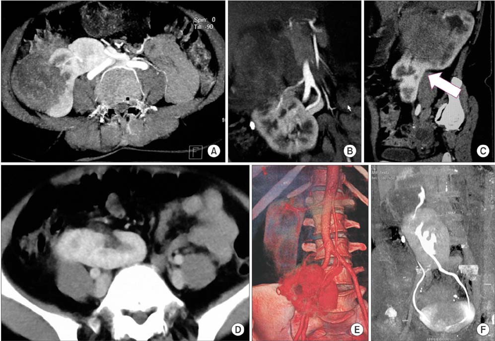
Fig. 1 (A-C) Axial, coronal and oblique sections show tumor arising from the orthotopic moiety. Arrow marks the junction between the ectopic and the orthotopic moiety. (D) Axial section shows horizontally lying pelvic ectopic kidney. (E) Three-dimensional angiogram demonstrating the origin and course of multiple renal arteries. (F) Delayed phase image depicting the pelvi-calyceal system and ureteric course of both moieties. The ureter of orthotopic moiety is coursing between tumor and ectopic moiety.
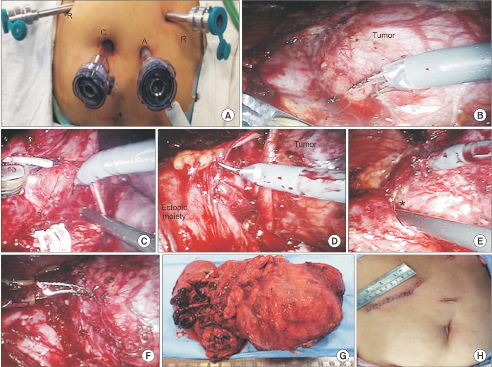
Fig. 2 Port placement (A); tumor being dissected (B); vascular pedicles being clipped and divided (C); ureter of orthotopic moiety dissected (D); plane of dissection between tumor mass and ectopic moiety (*) (E); tumor mass dissected all-around (F); gross specimen (G); postoperative wound (H). R, robotic arms; C, camera port, A, assistant port.
Reference
-
1. Shapiro E, Bauer SB, Chow JS. Anomalies of the upper urinary tract. In : Wein AJ, Kavoussi LR, Novick AC, Partin AW, Peters CA, editors. Campbell-Walsh urology. 10th ed. Philadelphia: Saunders;2012. p. 3140–3145.2. Stanley KE, Winfield HN, Donovan JF, Fallon B. Laparoscopic nephrectomy in crossed fused renal ectopia. Urology. 1993; 42:375–378.3. Felzenberg J, Nasrallah PF. Crossed renal ectopia without fusion associated with hydronephrosis in an infant. Urology. 1991; 38:450–452.4. Romero FR, Chan DY, Muntener M, Bagga HS, Brito FA, Kavoussi LR. Laparoscopic heminephrectomy for renal cell carcinoma in cross-fused ectopic kidney. Urology. 2007; 69:779.e11–779.e13.5. Stimac G, Dimanovski J, Ruzic B, Spajic B, Kraus O. Tumors in kidney fusion anomalies--report of five cases and review of the literature. Scand J Urol Nephrol. 2004; 38:485–489.6. Gur U, Yossepowitch O, Baniel J. Transitional cell carcinoma in a fused crossed ectopic kidney. Urology. 2003; 62:748.
