Giant Brunner’s Gland Hamartoma of the Duodenal Bulb Presenting with Upper Gastrointestinal Bleeding and Obstruction
- Affiliations
-
- 1Division of Gastroenterology, Department of Internal Medicine, Inje University Haeundae Paik Hospital, Inje University College of Medicine, Busan, Korea. ysydrim@gmail.com
- KMID: 2383455
- DOI: http://doi.org/10.5946/ce.2016.022
Abstract
- Brunner's gland hamartomas are small benign lesions that are most commonly found in the bulb of the duodenum. They are very uncommon, and most are found incidentally during upper gastrointestinal series or esophagogastroduodenoscopy. The lesions tend to be asymptomatic, but patients may present with symptoms of duodenal obstruction or hemorrhage secondary to ulceration. Histologically, a Brunner's gland hamartoma consists of the components of Brunner's gland cells, as well as glandular, adipose and muscle cells. In this study, we report the case of a 30-year-old man who presented with upper gastrointestinal bleeding and obstructive symptoms due to a giant Brunner's gland hamartoma in the duodenal bulb. The hamartoma was successfully removed by endoscopic resection. No significant complications were observed. Microscopically, the lesion was found to be entirely composed of variable Brunner's glands and adipocytes.
MeSH Terms
Figure
-
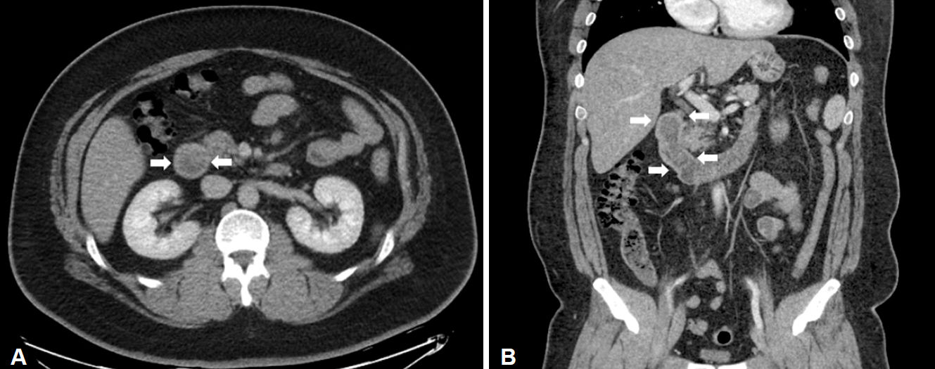
Fig. 1. (A) Contrast-enhanced axial and (B) coronal computed tomography scan shows a large polypoid mass in the duodenal bulb extending to second part of the duodenum (arrows).
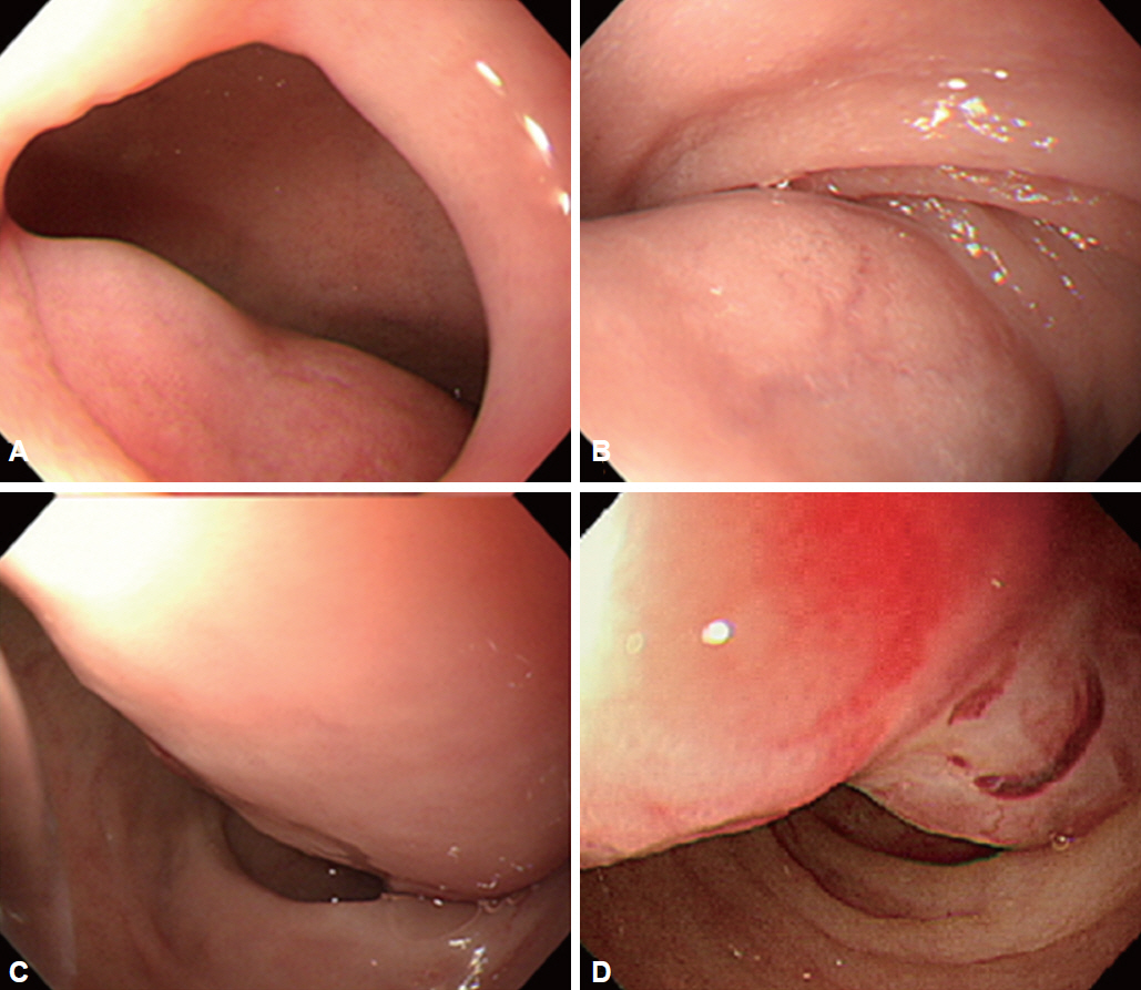
Fig. 2. Esophagogastroduodenoscopy shows a tortuous pedunculated mass occupying the lumen of the duodenal bulb and the second part of the duodenum. (A) Base of the tumor. (B) Thick trunk of the tumor at duodenal bulb. (C) Thick trunk at superior duodenal angle. (D) Tip of the tumor at the second part of the duodenum.
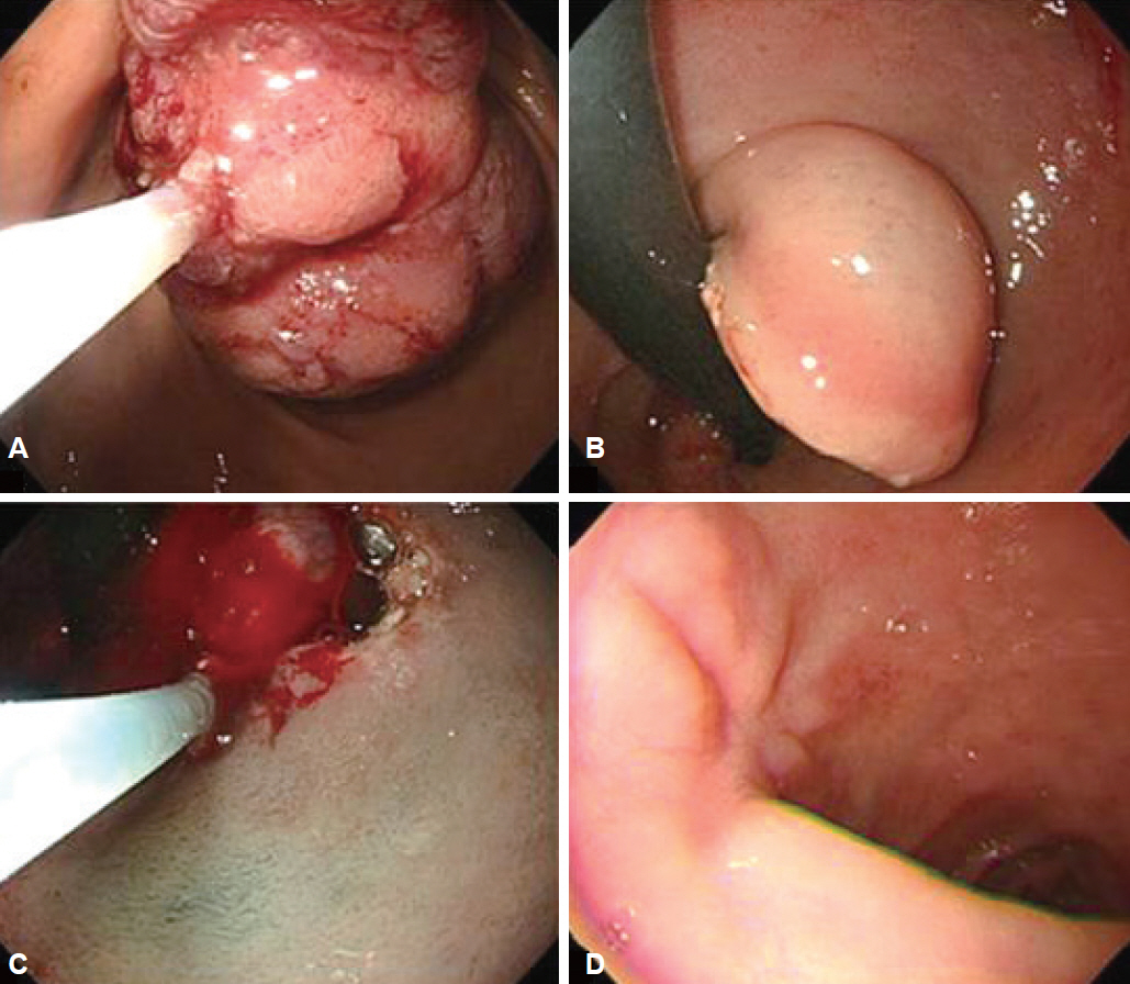
Fig. 3. Endoscopic removal. This shows the polyp being cut by two partial snare polypectomies and endoscopic submucosal dissection. It is successfully resected without any complications such as bleeding or perforation. (A) Stomach side. (B) Duodenal U-turn view. (C) Endoscopic submucosal dissection after U-turn. (D) Follow-up 2 months later.
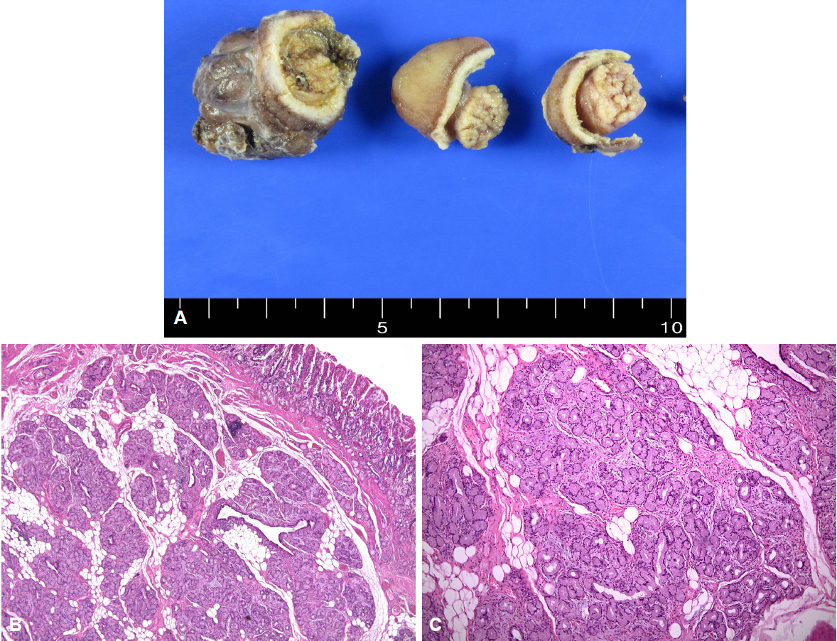
Fig. 4. Gross findings after formalin fixation. (A). A well-defined heterogenous yellow-white solid duodenal mass can be observed on the specimen section. The gross endoscopic resection specimen showed a large duodenal lesion measuring 9.3×2 cm. (B) Microscopic findings. Light microscopy revealed hyperplastic lobules of proliferating Brunner's glands separated by fibrous septum. (H&E stain, ×40). (C) Brunner's gland hyperplasia composed of variable size of Brunner’s glands (H&E stain, ×100) can be observed.
Reference
-
1. Nakabori T, Shinzaki S, Yamada T, et al. Atypical duodenal ulcer and invagination caused by a large pedunculated duodenal Brunner’s gland hamartoma. Gastrointest Endosc. 2014; 79:679–680.
Article2. Jung Y, Chung IK, Lee TH, et al. Successful endoscopic resection of large pedunculated brunner’s gland hamartoma causing gastrointestinal bleeding arising from the pylorus. Case Rep Gastroenterol. 2013; 7:304–307.
Article3. Sen R, Gupta V, Sharma N, Chawla N, Kumar S, Malik S. Brunner gland hamartoma masquerading as malignancy: a rare case report. Middle East J Dig Dis. 2014; 6:237–240.4. Gaspar JP, Stelow EB, Wang AY. Approach to the endoscopic resection of duodenal lesions. World J Gastroenterol. 2016; 22:600–617.
Article5. Lu L, Li R, Zhang G, Zhao Z, Fu W, Li W. Brunner’s gland adenoma of duodenum: report of two cases. Int J Clin Exp Pathol. 2015; 8:7565–7569.6. Martinez MA, Zyromski NJ, Luz LP. An unusual case of large symptomatic Brunner’s gland adenoma: endoscopic ultrasound imaging. Endosc Ultrasound. 2015; 4:266–267.
Article7. Cheung TT, Ip EW, Poon RT, Trendell-Smith N. Brunner’s gland adenoma: unusual cause of duodenal haemorrhage and obstruction. Hong Kong Med J. 2013; 19:460.e1–460.e2.
Article8. Stoos-Veic T, Tadic M, Aralica G. EUS-FNA of Brunner’s gland hamartoma: a case report. Cytopathology. 2013; 24:194–196.
Article
- Full Text Links
-
- Actions
-
Cited
- CITED
-
- Close
- Share
-
- Similar articles
-
- A Case of Giant Brunner's Gland Hamartoma Presenting as Gastrointestinal Hemorrhage
- Brunnera's Gland Hyperplasia: Treatment of Severe Diffuse Nodular Hyperplasia Mimicking a Malignancy on Pancreatic-Duodenal Area
- A Case of Brunner's Gland Hamartoma Presenting as Obscure Gastrointestinal Hemorrhage
- Endoscopic Resection of a Giant Duodenal Brunner's Gland Adenoma
- A Case of Hemorrhagic Angiodysplasia of the Duodenal Bulb in a Patient under Chronic Hemodialysis
