A Model of Glial Scarring Analogous to the Environment of a Traumatically Injured Spinal Cord Using Kainate
- Affiliations
-
- 1Department of Rehabilitation Medicine, Asan Medical Center, University of Ulsan College of Medicine, Seoul, Korea.
- 2Department of Physical Medicine and Rehabilitation, Ulsan University Hospital, University of Ulsan College of Medicine, Ulsan, Korea. chhwang1220ciba@gmail.com
- 3Department of Anatomy, Asan Medical Center, University of Ulsan College of Medicine, Seoul, Korea.
- KMID: 2382907
- DOI: http://doi.org/10.5535/arm.2016.40.5.757
Abstract
OBJECTIVE
To develop an in vitro model analogous to the environment of traumatic spinal cord injury (SCI), the authors evaluated change of astrogliosis following treatments with kainate and/or scratch, and degree of neurite outgrowth after treatment with a kainate inhibitor.
METHODS
Astrocytes were obtained from the rat spinal cord. Then, 99% of the cells were confirmed to be GFAP-positive astrocytes. For chemical injury, the cells were treated with kainate at different concentrations (10, 50 or 100 µM). For mechanical injury, two kinds of uniform scratches were made using a plastic pipette tip by removing strips of cells. For combined injury (S/K), scratch and kainate were provided. Cord neurons from rat embryos were plated onto culture plates immediately after the three kinds of injuries and some cultures were treated with a kainate inhibitor.
RESULTS
Astro-gliosis (glial fibrillary acidic protein [GFAP], vimentin, chondroitin sulfate proteoglycan [CSPG], rho-associated protein kinase [ROCK], and ephrin type-A receptor 4 [EphA4]) was most prominent after treatment with 50 µM kainate and extensive scratch injury in terms of single arm (p<0.001) and in the S/K-induced injury model in view of single or combination (p<0.001). Neurite outgrowth in the seeded spinal cord (β-III tubulin) was the least in the S/K-induced injury model (p<0.001) and this inhibition was reversed by the kainate inhibitor (p<0.001).
CONCLUSION
The current in vitro model combining scratch and kainate induced glial scarring and inhibitory molecules and restricted neurite outgrowth very strongly than either the mechanically or chemically-induced injury model; hence, it may be a useful tool for research on SCI.
MeSH Terms
Figure
-
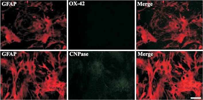
Fig. 1 Confirmation of depletion of microglia. Immunocytochemical staining of the culture system representing a pure astrocyte. GFAP, glial fibrillary acidic protein.
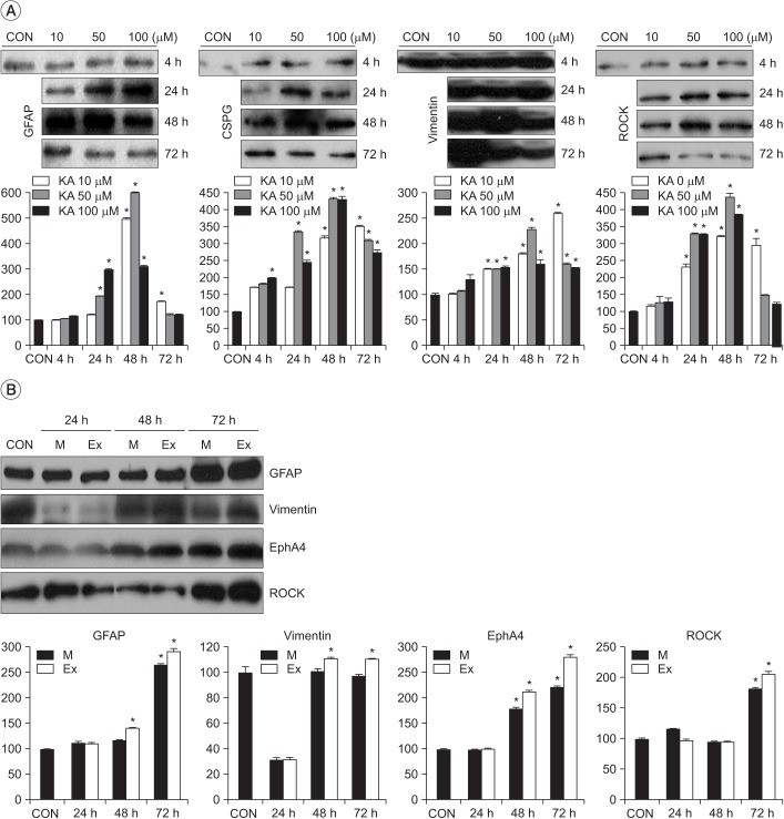
Fig. 2 Effects of injury intensity on scratch- and kainate-induced astrogliosis. (A) The optical immune-densities representing astrogliosis were the strongest at 50 µM kainate at 48 post-treatment hours. (B) The optical immune-densities representing astrogliosis were more prominent in the extensive (Ex) scratch injury model than in the moderate (M) scratch injury model. Values are presented as mean±standard deviation. GFAP, glial fibrillary acidic protein; CSPG, chondroitin sulfate proteoglycan; ROCK, rho-associated protein kinase; EphA4, ephrin type-A receptor 4; CON, control. *p<0.001 vs. CON; n=3–6.
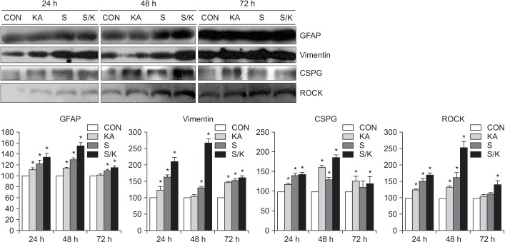
Fig. 3 Effects of single or combined injury on astrogliosis. The optical densities representing glia scarring were the strongest in the combined injury model among the control (CON), chemical (KA), mechanical (S), or combined injury (S/K) models and glial scarring in the S/K-induced injury model was the most intense at post-treatment 48 hours. Values are presented as mean±standard deviation. GFAP, glial fibrillary acidic protein; CSPG, chondroitin sulfate proteoglycan; ROCK, rho-associated protein kinase; KA, kainate; S, scratch; S/K, scratch/kainate. *p<0.001 vs. CON; n=3–6.
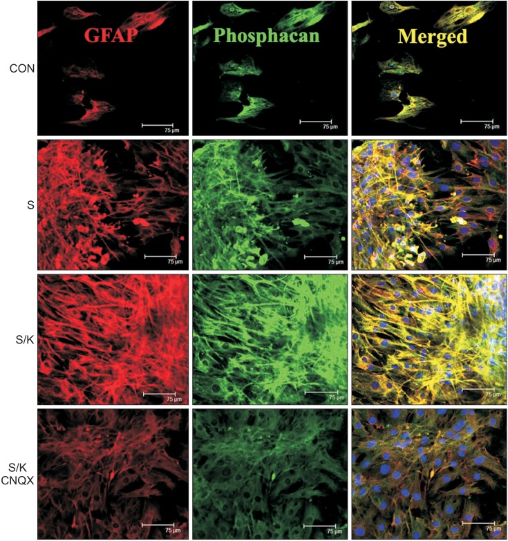
Fig. 4 Effects of kainate on expression of phosphacan (scale bar, 75 µm). Western blot was performed (n=3–6 for immunoblot analysis at each time point in each group). Stained GFAP and phosphacan representing astrogliosis were the largest in the scratch/kainate (S/K)-induced injury model among the control (CON), scratch (S), or S/K-induced injury models. Merged images (Merged) showed double-immunolabeling for GFAP and phosphacan. GFAP, glial fibrillary acidic protein; CNQX, 6-cyano-nitroquinoxaline-2,3-dione.
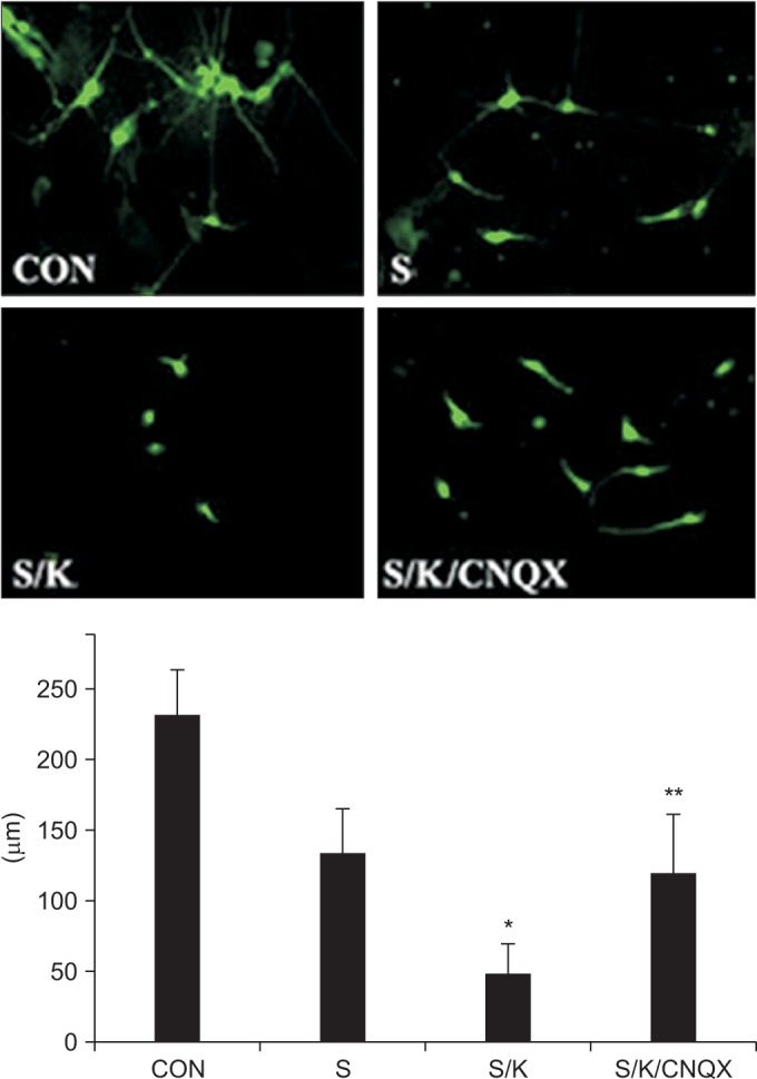
Fig. 5 Effects of the kainate inhibitor on neurite outgrowth. Neurite stained with anti-Tuj1 in the scratch/kainate (S/K)-induced injury model was the smallest among the control (CON), scratch (S), or S/K-induced injury models (*p<0.001 vs. S) and this effect was reversed by treatment with a kainate inhibitor (**p<0.001 vs. S/K).
Cited by 1 articles
-
Role of Agmatine on Neuroglia in Central Nervous System Injury
Sumit Barua, Jong Youl Kim, Jong Eun Lee
Brain Neurorehabil. 2019;12(1):. doi: 10.12786/bn.2019.12.e2.
Reference
-
1. Silver J, Miller JH. Regeneration beyond the glial scar. Nat Rev Neurosci. 2004; 5:146–156. PMID: 14735117.
Article2. McKeon RJ, Jurynec MJ, Buck CR. The chondroitin sulfate proteoglycans neurocan and phosphacan are expressed by reactive astrocytes in the chronic CNS glial scar. J Neurosci. 1999; 19:10778–10788. PMID: 10594061.
Article3. Hatten ME, Liem RK, Shelanski ML, Mason CA. Astroglia in CNS injury. Glia. 1991; 4:233–243. PMID: 1827781.
Article4. Yu AC, Lee YL, Eng LF. Astrogliosis in culture. I: The model and the effect of antisense oligonucleotides on glial fibrillary acidic protein synthesis. J Neurosci Res. 1993; 34:295–303. PMID: 8455207.
Article5. Calvo JL, Carbonell AL, Boya J. Co-expression of glial fibrillary acidic protein and vimentin in reactive astrocytes following brain injury in rats. Brain Res. 1991; 566:333–336. PMID: 1814551.6. Fronza M, Heinzmann B, Hamburger M, Laufer S, Merfort I. Determination of the wound healing effect of Calendula extracts using the scratch assay with 3T3 fibroblasts. J Ethnopharmacol. 2009; 126:463–467. PMID: 19781615.
Article7. Nishio T, Kawaguchi S, Yamamoto M, Iseda T, Kawasaki T, Hase T. Tenascin-C regulates proliferation and migration of cultured astrocytes in a scratch wound assay. Neuroscience. 2005; 132:87–102. PMID: 15780469.
Article8. Smith AN, Willis E, Chan VT, Muffley LA, Isik FF, Gibran NS, et al. Mesenchymal stem cells induce dermal fibroblast responses to injury. Exp Cell Res. 2010; 316:48–54. PMID: 19666021.
Article9. Park E, Velumian AA, Fehlings MG. The role of excitotoxicity in secondary mechanisms of spinal cord injury: a review with an emphasis on the implications for white matter degeneration. J Neurotrauma. 2004; 21:754–774. PMID: 15253803.
Article10. Matute C, Domercq M, Sanchez-Gomez MV. Glutamate-mediated glial injury: mechanisms and clinical importance. Glia. 2006; 53:212–224. PMID: 16206168.
Article11. Liu D, Xu GY, Pan E, McAdoo DJ. Neurotoxicity of glutamate at the concentration released upon spinal cord injury. Neuroscience. 1999; 93:1383–1389. PMID: 10501463.
Article12. Olney JW, Rhee V, Ho OL. Kainic acid: a powerful neurotoxic analogue of glutamate. Brain Res. 1974; 77:507–512. PMID: 4152936.
Article13. Mazzone GL, Margaryan G, Kuzhandaivel A, Nasrabady SE, Mladinic M, Nistri A. Kainate-induced delayed onset of excitotoxicity with functional loss unrelated to the extent of neuronal damage in the in vitro spinal cord. Neuroscience. 2010; 168:451–462. PMID: 20362644.
Article14. Yang H, Cheng XP, Li JW, Yao Q, Ju G. De-differentiation response of cultured astrocytes to injury induced by scratch or conditioned culture medium of scratch-insulted astrocytes. Cell Mol Neurobiol. 2009; 29:455–473. PMID: 19130217.
Article15. Yoshimura E, Majima A, Sakakura Y, Sakakura T, Yoshida T. Expression of tenascin-C and the integrin alpha 9 subunit in regeneration of rat nasal mucosa after chemical injury: involvement in migration and proliferation of epithelial cells. Histochem Cell Biol. 1999; 111:259–264. PMID: 10219625.16. O'Callaghan JP, Jensen KF, Miller DB. Quantitative aspects of drug and toxicant-induced astrogliosis. Neurochem Int. 1995; 26:115–124. PMID: 7599532.17. Ellis EF, McKinney JS, Willoughby KA, Liang S, Povlishock JT. A new model for rapid stretch-induced injury of cells in culture: characterization of the model using astrocytes. J Neurotrauma. 1995; 12:325–339. PMID: 7473807.
Article18. Milenkovic I, Nedeljkovic N, Filipovic R, Pekovic S, Culic M, Rakic L, et al. Pattern of glial fibrillary acidic protein expression following kainate-induced cerebellar lesion in rats. Neurochem Res. 2005; 30:207–213. PMID: 15895824.
Article19. David JC, Yamada KA, Bagwe MR, Goldberg MP. AMPA receptor activation is rapidly toxic to cortical astrocytes when desensitization is blocked. J Neurosci. 1996; 16:200–209. PMID: 8613786.
Article20. Malhotra SK, Luong LT, Bhatnagar R, Shnitka TK. Up-regulation of reactive astrogliosis in the rat glioma 9L cell line by combined mechanical and chemical injuries. Cytobios. 1997; 89:115–134. PMID: 9363621.21. Wanner IB, Deik A, Torres M, Rosendahl A, Neary JT, Lemmon VP, et al. A new in vitro model of the glial scar inhibits axon growth. Glia. 2008; 56:1691–1709. PMID: 18618667.22. Gaviria M, Privat A, d'Arbigny P, Kamenka JM, Haton H, Ohanna F. Neuroprotective effects of gacyclidine after experimental photochemical spinal cord lesion in adult rats: dose-window and time-window effects. J Neurotrauma. 2000; 17:19–30. PMID: 10674755.
Article23. Rosenberg LJ, Teng YD, Wrathall JR. 2,3-Dihydroxy-6-nitro-7-sulfamoyl-benzo(f)quinoxaline reduces glial loss and acute white matter pathology after experimental spinal cord contusion. J Neurosci. 1999; 19:464–475. PMID: 9870974.
Article24. Wrathall JR, Teng YD, Choiniere D. Amelioration of functional deficits from spinal cord trauma with systemically administered NBQX, an antagonist of non-N-methyl-D-aspartate receptors. Exp Neurol. 1996; 137:119–126. PMID: 8566203.
Article25. Chang ML, Wu CH, Jiang-Shieh YF, Shieh JY, Wen CY. Reactive changes of retinal astrocytes and Müller glial cells in kainate-induced neuroexcitotoxicity. J Anat. 2007; 210:54–65. PMID: 17229283.
Article26. Kohno H, Sakai T, Kitahara K. Induction of nestin, Ki-67, and cyclin D1 expression in Müller cells after laser injury in adult rat retina. Graefes Arch Clin Exp Ophthalmol. 2006; 244:90–95. PMID: 15983812.
Article27. Schnell L, Fearn S, Klassen H, Schwab ME, Perry VH. Acute inflammatory responses to mechanical lesions in the CNS: differences between brain and spinal cord. Eur J Neurosci. 1999; 11:3648–3658. PMID: 10564372.
Article28. McKeon RJ, Schreiber RC, Rudge JS, Silver J. Reduction of neurite outgrowth in a model of glial scarring following CNS injury is correlated with the expression of inhibitory molecules on reactive astrocytes. J Neurosci. 1991; 11:3398–3411. PMID: 1719160.
Article29. Maxwell WL, Follows R, Ashhurst DE, Berry M. The response of the cerebral hemisphere of the rat to injury. II: The neonatal rat. Philos Trans R Soc Lond B Biol Sci. 1990; 328:501–513. PMID: 1974075.30. Shearer MC, Niclou SP, Brown D, Asher RA, Holtmaat AJ, Levine JM, et al. The astrocyte/meningeal cell interface is a barrier to neurite outgrowth which can be overcome by manipulation of inhibitory molecules or axonal signalling pathways. Mol Cell Neurosci. 2003; 24:913–925. PMID: 14697658.
Article31. Choi DW. Glutamate neurotoxicity and diseases of the nervous system. Neuron. 1988; 1:623–634. PMID: 2908446.
Article32. Akins PT, Atkinson RP. Glutamate AMPA receptor antagonist treatment for ischaemic stroke. Curr Med Res Opin. 2002; 18(Suppl 2):s9–s13. PMID: 12365832.
Article33. Lerma J. Roles and rules of kainate receptors in synaptic transmission. Nat Rev Neurosci. 2003; 4:481–495. PMID: 12778120.
Article34. Jones LL, Sajed D, Tuszynski MH. Axonal regeneration through regions of chondroitin sulfate proteoglycan deposition after spinal cord injury: a balance of permissiveness and inhibition. J Neurosci. 2003; 23:9276–9288. PMID: 14561854.
Article35. Hansson E, Muyderman H, Leonova J, Allansson L, Sinclair J, Blomstrand F, et al. Astroglia and glutamate in physiology and pathology: aspects on glutamate transport, glutamate-induced cell swelling and gap-junction communication. Neurochem Int. 2000; 37:317–329. PMID: 10812217.
Article36. Gottlieb M, Matute C. Expression of ionotropic glutamate receptor subunits in glial cells of the hippocampal CA1 area following transient forebrain ischemia. J Cereb Blood Flow Metab. 1997; 17:290–300. PMID: 9119902.
Article37. Simantov R, Crispino M, Hoe W, Broutman G, Tocco G, Rothstein JD, et al. Changes in expression of neuronal and glial glutamate transporters in rat hippocampus following kainate-induced seizure activity. Brain Res Mol Brain Res. 1999; 65:112–123. PMID: 10036313.
Article38. Aono S, Kashiwamata S. GFAP under physiological and pathological conditions. Med Sci Res. 1990; 18:235–239.
- Full Text Links
-
- Actions
-
Cited
- CITED
-
- Close
- Share
-
- Similar articles
-
- Effects of Fetal Spinal Cord Transplants on Injured Rat Spinal Cord
- Fracture of Femur Neck with Heterotopic Ossification in Spinal Cord Injured Patient
- Olig2-expressing Mesenchymal Stem Cells Enhance Functional Recovery after Contusive Spinal Cord Injury
- Activation of Embryonic Intermediate Filaments Contributes to Glial Scar Formation after Spinal Cord Injury in Rats
- The Influence of Self-care Agency and Social Support on Self-care Practice among Spinal Cord Injured Patients
