J Pathol Transl Med.
2015 Sep;49(5):403-408. 10.4132/jptm.2015.05.28.
Paediatric Primary Pachymeningeal Xanthogranuloma with Scattered Foci Displaying Reticulohistiocytoma-like Features
- Affiliations
-
- 1Anatomical Pathology Division, "Dr. Manuel Gea Gonzalez" General Hospital, Mexico City, Mexico. k7nigricans@hotmail.com
- 2Pathology Unit, Neuropathology Service, Mexico General Hospital, Mexico City, Mexico.
- 3Paediatric Neurosurgery, "Legaria" Paediatric Hospital, Mexico City, Mexico.
- KMID: 2381396
- DOI: http://doi.org/10.4132/jptm.2015.05.28
Abstract
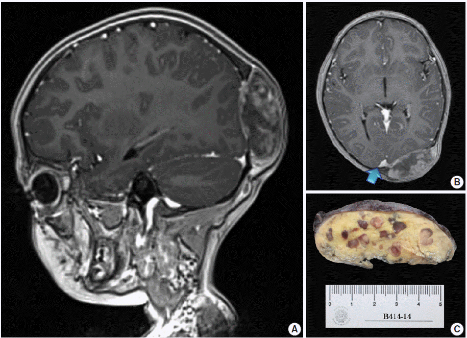
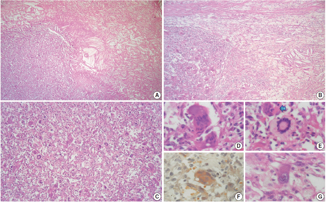
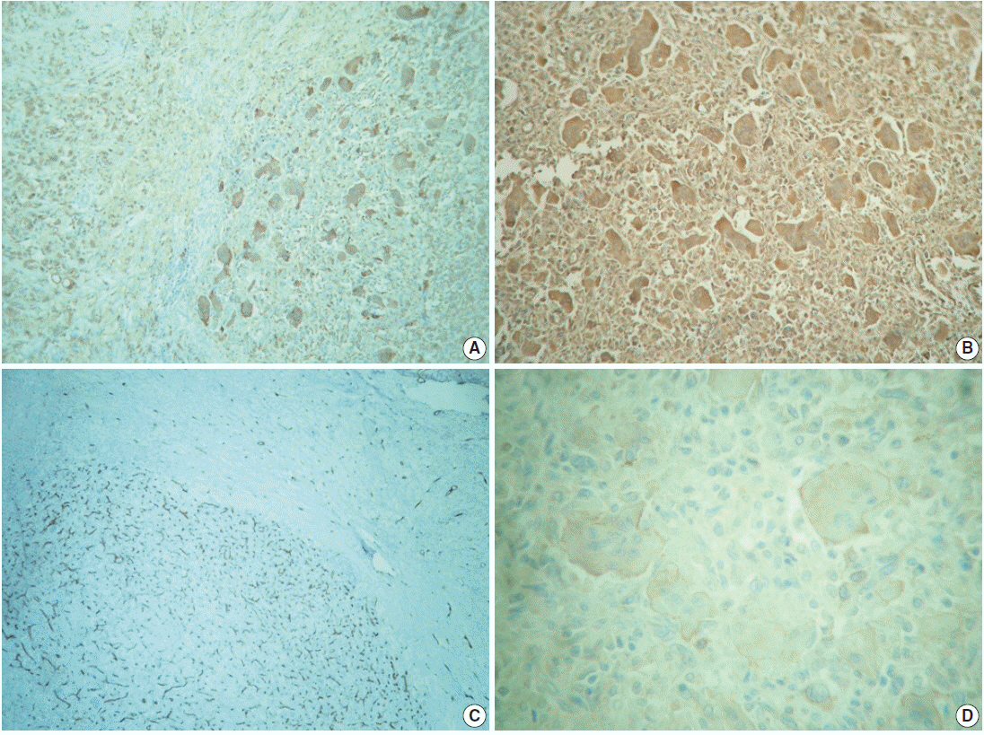
- We report a unique case of a 4-year-old girl with an intriguing fibrohistiocytic tumour. Magnetic resonance imaging scans showed a dural mass of variegated intensity compressing the left occipital pole and apparently extending toward the superior sagittal sinus. Grossly, the cut surface of the surgical specimen was yellow, pale, and soft with reddish kernel-like crusts. Histologically, the yellow areas resembled cholesterol granulomas with widespread coagulative necrosis, cholesterol clefts, powdery calcification, foreign body-type giant cells, and foamy macrophages, while the scattered red spots contained numerous multinucleated giant cells of foreign-body and Touton types, the former with amphophilic to slightly eosinophilic cytoplasm. Immunoperoxidase reactions confirmed the expression of histiocytic markers and vimentin. As far as we know, no tumour displaying these peculiar morphological features has yet been described.
Keyword
MeSH Terms
Figure
Reference
-
1. Goldblum JR, Folpe AL, Weiss SW. Benign fibrohistiocytic and histiocytic tumors. In : Goldblum JR, Folpe AL, Weiss SW, editors. Enzinger and Weiss’s soft tissue tumors. 6th ed. Philadelphia: Elsevier-Saunders;2014. p. 341–86.2. Paulus W, Perry A. Histiocytic tumours. In : Louis DN, Ohgaki H, Wiestler OD, editors. WHO classification of tumours of the central nervous system. 4th ed. Lyon: IARC Press;2007. p. 193–6.3. Black J, Coffin CM, Dehner LP. Fibrohistiocytic tumors and related neoplasms in children and adolescents. Pediatr Dev Pathol. 2012; 15(1 Suppl):181–210.
Article4. Chung IH, Lee YS, Myong NH, Lee MJ, Lee SK, Ko JH. Intracranial fibroxanthoma in an infant: a case report. Korean J Radiol. 2009; 10:402–6.
Article5. Kim DS, Kim TS, Choi JU. Intradural extramedullary xanthoma of the spine: a rare lesion arising from the dura mater of the spine: case report. Neurosurgery. 1996; 39:182–5.
Article6. Miyazono M, Nishio S, Morioka T, Hamada Y, Fukui M, Yanai S. Fibroxanthoma arising from the cranial dura mater. Neurosurg Rev. 1999; 22:215–8.
Article7. Usul H, Kuzeyli K, Cakir E, et al. Giant cranial extradural primary fibroxanthoma: a case report. Surg Neurol. 2005; 63:281–4.
Article8. Pimentel J, Fernandes A, Távora L, Miguéns J, Lobo Antunes J. Benign isolated fibrohistiocytic tumor arising from the central nervous system: considerations about two cases. Clin Neuropathol. 2002; 21:93–8.9. Harries AM, Mitchell R. Haemorrhagic cerebellar fibrous histiocytoma: case report and literature review. Br J Neurosurg. 2011; 25:120–1.
Article10. Kepes JJ, Kepes M, Slowik F. Fibrous xanthomas and xanthosarcomas of the meninges and the brain. Acta Neuropathol. 1973; 23:187–99.
Article11. Blumer G. Bilateral cholesteatomatous endothelioma of the choroid plexus. Johns Hopkins Hosp Rep. 1900; 9:279–90.12. Lam RM, Colah SA. Atypical fibrous histiocytoma with myxoid stroma: a rare lesion arising from dura mater of the brain. Cancer. 1979; 43:237–45.13. Pear BL. Xanthogranuloma of the choroid plexus. AJR Am J Roentgenol. 1984; 143:401–2.
Article14. Berti E, Zelger B, Caputo R. Reticulohistiocytosis. In : LeBoit PE, Burg G, Weedon D, Sarasin A, editors. World Health Organization classification of tumours: pathology and genetics of skin tumours. Lyon: IARC Press;2006. p. 224–5.15. Miettinen M, Fetsch JF. Reticulohistiocytoma (solitary epithelioid histiocytoma): a clinicopathologic and immunohistochemical study of 44 cases. Am J Surg Pathol. 2006; 30:521–8.16. Muenchau A, Laas R. Xanthogranuloma and xanthoma of the choroid plexus: evidence for different etiology and pathogenesis. Clin Neuropathol. 1997; 16:72–6.17. Perry A, Dehner LP. Meningeal tumors of childhood and infancy. An update and literature review. Brain Pathol. 2003; 13:386–408.
Article18. Sarkar C, Roy S, Bhatia S. Xanthomatous change in tumours of glial origin. Indian J Med Res. 1990; 92:324–31.19. Leong CF, Raudhawati O, Cheong SK, Sivagengei K, Noor Hamidah H. Epithelial membrane antigen (EMA) or MUC1 expression in monocytes and monoblasts. Pathology. 2003; 35:422–7.
Article20. Ozzello L, Stout AP, Murray MR. Cultural characteristics of malignant histiocytomas and fibrous xanthomas. Cancer. 1963; 16:331–44.
Article


