Advances in the Endoscopic Assessment of Inflammatory Bowel Diseases: Cooperation between Endoscopic and Pathologic Evaluations
- Affiliations
-
- 1Department of Internal Medicine, Yonsei University College of Medicine, Seoul, Korea. geniushee@yuhs.ac
- KMID: 2381379
- DOI: http://doi.org/10.4132/jptm.2015.04.09
Abstract
- Endoscopic assessment has a crucial role in the management of inflammatory bowel disease (IBD). It is particularly useful for the assessment of IBD disease extension, severity, and neoplasia surveillance. Recent advances in endoscopic imaging techniques have been revolutionized over the past decades, progressing from conventional white light endoscopy to novel endoscopic techniques using molecular probes or electronic filter technologies. These new technologies allow for visualization of the mucosa in detail and monitor for inflammation/dysplasia at the cellular or sub-cellular level. These techniques may enable us to alter the IBD surveillance paradigm from four quadrant random biopsy to targeted biopsy and diagnosis. High definition endoscopy and dye-based chromoendoscopy can improve the detection rate of dysplasia and evaluate inflammatory changes with better visualization. Dye-less chromoendoscopy, including narrow band imaging, iScan, and autofluorescence imaging can also enhance surveillance in comparison to white light endoscopy with optical or electronic filter technologies. Moreover, confocal laser endomicroscopy or endocytoscopy have can achieve real-time histology evaluation in vivo and have greater accuracy in comparison with histology. These new technologies could be combined with standard endoscopy or further histologic confirmation in patients with IBD. This review offers an evidence-based overview of new endoscopic techniques in patients with IBD.
Keyword
MeSH Terms
Figure
-
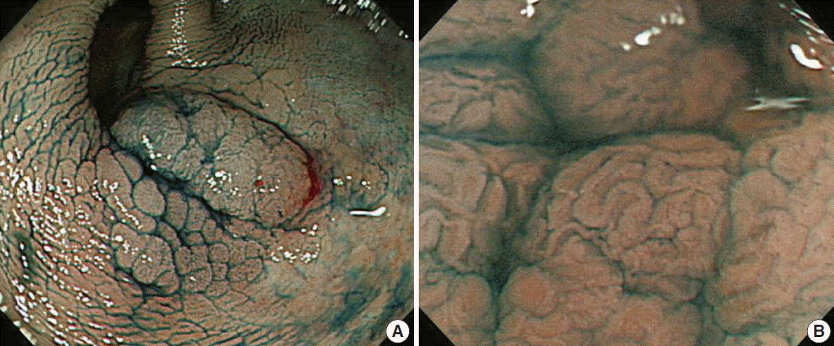
Fig. 1. Chromoendoscopy using indigocarmine (A) and combined with magnification technique (B) for colonic dysplasia in ulcerative colitis (Courtesy of Dr. Jeong-Sik Byeon at Asan Medical Center).
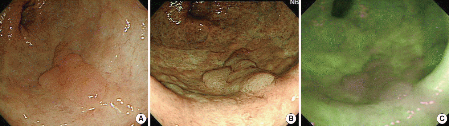
Fig. 2. Observation findings of colonic dysplasia using white light endoscopy (A), narrow band imaging technique (B), and autofluorescence imaging technique (C) in ulcerative colitis (Courtesy of Dr. Jeong-Sik Byeon at Asan Medical Center).
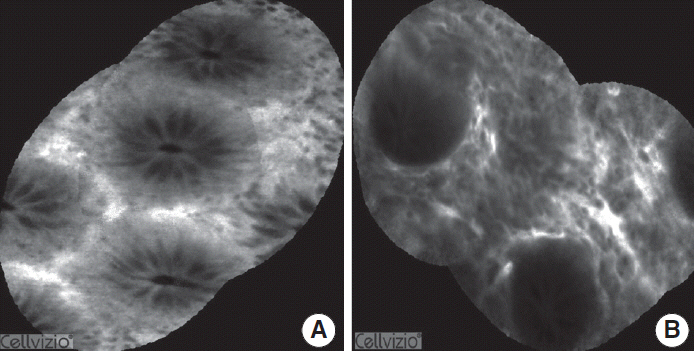
Fig. 3. Confocal laser endomicroscopic findings for normal mucosa (A) and mucosa in active ulcerative colitis (B). In ulcerative colitis, lamina propria widening, inflammatory infiltrates, goblet cell depletion, and crypt distortion are observed.
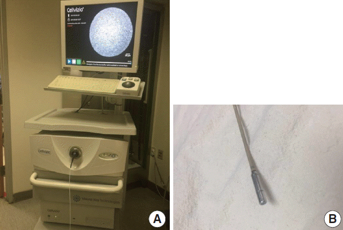
Fig. 4. Cellvizio system for probe based confocal laser endomicroscopy (A) and a probe (B).
Cited by 1 articles
-
Probe-based confocal laser endomicroscopy in the differential diagnosis of inflammatory bowel diseases: a case series
Jung Won Park, Tae Il Kim, Jae Hee Cheon
Intest Res. 2018;16(4):641-645. doi: 10.5217/ir.2018.00035.
Reference
-
1. Hommes DW, van Deventer SJ. Endoscopy in inflammatory bowel diseases. Gastroenterology. 2004; 126:1561–73.
Article2. Stange EF, Travis SP, Vermeire S, et al. European evidence based consensus on the diagnosis and management of Crohn’s disease: definitions and diagnosis. Gut. 2006; 55 Suppl 1:i1–15.
Article3. Rubin DC, Shaker A, Levin MS. Chronic intestinal inflammation: inflammatory bowel disease and colitis-associated colon cancer. Front Immunol. 2012; 3:107.
Article4. Eaden JA, Abrams KR, Mayberry JF. The risk of colorectal cancer in ulcerative colitis: a meta-analysis. Gut. 2001; 48:526–35.
Article5. Laukoetter MG, Mennigen R, Hannig CM, et al. Intestinal cancer risk in Crohn’s disease: a meta-analysis. J Gastrointest Surg. 2011; 15:576–83.
Article6. Jess T, Simonsen J, Jørgensen KT, Pedersen BV, Nielsen NM, Frisch M, et al. Decreasing risk of colorectal cancer in patients with inflammatory bowel disease over 30 years. Gastroenterology. 2012; 143:375–81. e1.
Article7. Cheon JH, Kim WH. Recent advances of endoscopy in inflammatory bowel diseases. Gut Liver. 2007; 1:118–25.
Article8. Rutter MD, Saunders BP, Wilkinson KH, et al. Thirty-year analysis of a colonoscopic surveillance program for neoplasia in ulcerative colitis. Gastroenterology. 2006; 130:1030–8.
Article9. Panaccione R. The approach to dysplasia surveillance in inflammatory bowel disease. Can J Gastroenterol. 2006; 20:251–3.
Article10. Biancone L, Michetti P, Travis S, et al. European evidence-based consensus on the management of ulcerative colitis: special situations. J Crohns Colitis. 2008; 2:63–92.
Article11. Eaden JA, Mayberry JF; British Society for Gastroenterology; Association of Coloproctology for Great Britain and Ireland. Guidelines for screening and surveillance of asymptomatic colorectal cancer in patients with inflammatory bowel disease. Gut. 2002; 51 Suppl 5:V10–2.
Article12. Kiesslich R, Goetz M, Lammersdorf K, et al. Chromoscopy-guided endomicroscopy increases the diagnostic yield of intraepithelial neoplasia in ulcerative colitis. Gastroenterology. 2007; 132:874–82.
Article13. Hlavaty T, Huorka M, Koller T, et al. Colorectal cancer screening in patients with ulcerative and Crohn’s colitis with use of colonoscopy, chromoendoscopy and confocal endomicroscopy. Eur J Gastroenterol Hepatol. 2011; 23:680–9.
Article14. ASGE Technology Committee. High-definition and high-magnification endoscopes. Gastrointest Endosc. 2014; 80:919–27.15. Udagawa T, Amano M, Okada F. Development of magnifying video endoscopes with high resolution. Dig Endosc. 2001; 13:163–9.
Article16. Subramanian V, Ramappa V, Telakis E, et al. Comparison of high definition with standard white light endoscopy for detection of dysplastic lesions during surveillance colonoscopy in patients with colonic inflammatory bowel disease. Inflamm Bowel Dis. 2013; 19:350–5.
Article17. Kiesslich R, Neurath MF. Chromoendoscopy in inflammatory bowel disease. Gastroenterol Clin North Am. 2012; 41:291–302.
Article18. Canto MI. Staining in gastrointestinal endoscopy: the basics. Endoscopy. 1999; 31:479–86.
Article19. Subramanian V, Mannath J, Ragunath K, Hawkey CJ. Meta-analysis: the diagnostic yield of chromoendoscopy for detecting dysplasia in patients with colonic inflammatory bowel disease. Aliment Pharmacol Ther. 2011; 33:304–12.
Article20. Soetikno R, Subramanian V, Kaltenbach T, et al. The detection of nonpolypoid (flat and depressed) colorectal neoplasms in patients with inflammatory bowel disease. Gastroenterology. 2013; 144:1349–52. e1-6.
Article21. Goetz M, Kiesslich R. Advanced imaging of the gastrointestinal tract: research vs. clinical tools? Curr Opin Gastroenterol. 2009; 25:412–21.
Article22. Amateau SK, Canto MI. Enhanced mucosal imaging. Curr Opin Gastroenterol. 2010; 26:445–52.
Article23. Kuznetsov K, Lambert R, Rey JF. Narrow-band imaging: potential and limitations. Endoscopy. 2006; 38:76–81.
Article24. Subramanian V, Bisschops R. Image-enhanced endoscopy is critical in the surveillance of patients with colonic IBD. Gastrointest Endosc Clin N Am. 2014; 24:393–403.
Article25. Tontini GE, Vecchi M, Neurath MF, Neumann H. Review article: newer optical and digital chromoendoscopy techniques vs. dyebased chromoendoscopy for diagnosis and surveillance in inflammatory bowel disease. Aliment Pharmacol Ther. 2013; 38:1198–208.
Article26. van den Broek FJ, Reitsma JB, Curvers WL, Fockens P, Dekker E. Systematic review of narrow-band imaging for the detection and differentiation of neoplastic and nonneoplastic lesions in the colon (with videos). Gastrointest Endosc. 2009; 69:124–35.
Article27. Pohl J, Nguyen-Tat M, Pech O, May A, Rabenstein T, Ell C. Computed virtual chromoendoscopy for classification of small colorectal lesions: a prospective comparative study. Am J Gastroenterol. 2008; 103:562–9.
Article28. Teixeira CR, Torresini RS, Canali C, et al. Endoscopic classification of the capillary-vessel pattern of colorectal lesions by spectral estimation technology and magnifying zoom imaging. Gastrointest Endosc. 2009; 69(3 Pt 2):750–6.
Article29. East JE, Suzuki N, von Herbay A, Saunders BP. Narrow band imaging with magnification for dysplasia detection and pit pattern assessment in ulcerative colitis surveillance: a case with multiple dysplasia associated lesions or masses. Gut. 2006; 55:1432–5.
Article30. Dekker E, van den Broek FJ, Reitsma JB, et al. Narrow-band imaging compared with conventional colonoscopy for the detection of dysplasia in patients with longstanding ulcerative colitis. Endoscopy. 2007; 39:216–21.
Article31. Ignjatovic A, East JE, Subramanian V, et al. Narrow band imaging for detection of dysplasia in colitis: a randomized controlled trial. Am J Gastroenterol. 2012; 107:885–90.
Article32. van den Broek FJ, Fockens P, van Eeden S, et al. Narrow-band imaging versus high-definition endoscopy for the diagnosis of neoplasia in ulcerative colitis. Endoscopy. 2011; 43:108–15.
Article33. Pellisé M, López-Cerón M, Rodríguez de Miguel C, et al. Narrowband imaging as an alternative to chromoendoscopy for the detection of dysplasia in long-standing inflammatory bowel disease: a prospective, randomized, crossover study. Gastrointest Endosc. 2011; 74:840–8.
Article34. Hoffman A, Sar F, Goetz M, et al. High definition colonoscopy combined with i-Scan is superior in the detection of colorectal neoplasias compared with standard video colonoscopy: a prospective randomized controlled trial. Endoscopy. 2010; 42:827–33.
Article35. Kodashima S, Fujishiro M. Novel image-enhanced endoscopy with i-scan technology. World J Gastroenterol. 2010; 16:1043–9.
Article36. Neumann H, Vieth M, Günther C, et al. Virtual chromoendoscopy for prediction of severity and disease extent in patients with inflammatory bowel disease: a randomized controlled study. Inflamm Bowel Dis. 2013; 19:1935–42.37. Neumann H, Vieth M, Tontini GE, et al. 33 High-definition endoscopy with virtual chromoendoscopy for prediction of food allergy in real-time: a prospective, randomized study with cross-over design. Gastroenterology. 2014; 146(5 Suppl 1):S-9–S-10.
Article38. Liu JT, Loewke NO, Mandella MJ, Levenson RM, Crawford JM, Contag CH. Point-of-care pathology with miniature microscopes. Anal Cell Pathol (Amst). 2011; 34:81–98.
Article39. Dunbar K, Canto M. Confocal endomicroscopy. Curr Opin Gastroenterol. 2008; 24:631–7.
Article40. Iacucci M, Panaccione R, Ghosh S. Advances in novel diagnostic endoscopic imaging techniques in inflammatory bowel disease. Inflamm Bowel Dis. 2013; 19:873–80.
Article41. Watanabe O, Ando T, Maeda O, et al. Confocal endomicroscopy in patients with ulcerative colitis. J Gastroenterol Hepatol. 2008; 23 Suppl 2:S286–90.
Article42. Li CQ, Xie XJ, Yu T, et al. Classification of inflammation activity in ulcerative colitis by confocal laser endomicroscopy. Am J Gastroenterol. 2010; 105:1391–6.
Article43. Kiesslich R, Burg J, Vieth M, et al. Confocal laser endoscopy for diagnosing intraepithelial neoplasias and colorectal cancer in vivo. Gastroenterology. 2004; 127:706–13.44. Li CQ, Liu J, Ji R, Li Z, Xie XJ, Li YQ. Use of confocal laser endomicroscopy to predict relapse of ulcerative colitis. BMC Gastroenterol. 2014; 14:45.
Article45. Watanabe C, Sumioka M, Hiramoto T, et al. Magnifying colonoscopy used to predict disease relapse in patients with quiescent ulcerative colitis. Inflamm Bowel Dis. 2009; 15:1663–9.
Article46. Neumann H, Vieth M, Atreya R, et al. Assessment of Crohn’s disease activity by confocal laser endomicroscopy. Inflamm Bowel Dis. 2012; 18:2261–9.
Article47. Asaoka D, Nagahara A, Kurosawa A, et al. Utility of autofluorescence imaging videoendoscopy system for the detection of minimal changes associated with reflux esophagitis. Endoscopy. 2008; 40 Suppl 2:E172–3.
Article48. Inoue T, Uedo N, Ishihara R, et al. Autofluorescence imaging videoendoscopy in the diagnosis of chronic atrophic fundal gastritis. J Gastroenterol. 2010; 45:45–51.
Article49. Asaoka D, Nagahara A, Oguro M, et al. Utility of autofluorescence imaging videoendoscopy in screening for Barrett’s esophagus. Endoscopy. 2009; 41 Suppl 2:E113.
Article50. Nakaniwa N, Namihisa A, Ogihara T, et al. Newly developed autofluorescence imaging videoscope system for the detection of colonic neoplasms. Dig Endosc. 2005; 17:235–40.
Article51. van den Broek FJ, Fockens P, van Eeden S, et al. Endoscopic trimodal imaging for surveillance in ulcerative colitis: randomised comparison of high-resolution endoscopy and autofluorescence imaging for neoplasia detection; and evaluation of narrow-band imaging for classification of lesions. Gut. 2008; 57:1083–9.
Article52. Osada T, Arakawa A, Sakamoto N, et al. Autofluorescence imaging endoscopy for identification and assessment of inflammatory ulcerative colitis. World J Gastroenterol. 2011; 17:5110–6.
Article53. Neumann H, Vieth M, Neurath MF. Image of the month. Endocytoscopy-based detection of focal high-grade intraepithelial neoplasia in colonic polyps. Clin Gastroenterol Hepatol. 2011; 9:e13.54. Neumann H, Vieth M, Neurath MF, Atreya R. Endocytoscopy allows accurate in vivo differentiation of mucosal inflammatory cells in IBD: a pilot study. Inflamm Bowel Dis. 2013; 19:356–62.55. Pohl H, Rosch T, Tanczos BT, Rudolph B, Schlüns K, Baumgart DC. Endocytoscopy for the detection of microstructural features in adult patients with celiac sprue: a prospective, blinded endocytoscopyconventional histology correlation study. Gastrointest Endosc. 2009; 70:933–41.
Article56. Cipolletta L, Bianco MA, Rotondano G, et al. Endocytoscopy can identify dysplasia in aberrant crypt foci of the colorectum: a prospective in vivo study. Endoscopy. 2009; 41:129–32.
- Full Text Links
-
- Actions
-
Cited
- CITED
-
- Close
- Share
-
- Similar articles
-
- Diagnostic Tips for Making the Diagnosis of Inflammatory Bowel Disease
- Endoscopic activity in inflammatory bowel disease: clinical significance and application in practice
- Small Bowel Endoscopy in Inflammatory Bowel Disease
- Endoscopic molecular imaging in inflammatory bowel disease
- Endoscopic Diagnosis and Differentiation of Inflammatory Bowel Disease
