J Korean Neurosurg Soc.
2017 Feb;60(2):174-180. 10.3340/jkns.2016.0707.014.
Risk Factors of Proximal Junctional Kyphosis after Multilevel Fusion Surgery: More Than 2 Years Follow-Up Data
- Affiliations
-
- 1Department of Neurosurgery, Inha University Hospital, Inha University School of Medicine, Incheon, Korea. nsyoon@gmail.com
- 2Department of Neurosurgery, Na Eun Hospital, Incheon, Korea.
- 3Ba Reun Na Mu Neurosurgical Clinic, Incheon, Korea.
- 4Department of Neurosurgery, Asan Medical Center, University of Ulsan College of Medicine, Seoul, Korea.
- KMID: 2374878
- DOI: http://doi.org/10.3340/jkns.2016.0707.014
Abstract
OBJECTIVE
Proximal junctional kyphosis (PJK) is radiologic finding, and is defined as kyphosis of >10° at the proximal end of a construct. The aim of this study is to identify factors associated with PJK after segmental spinal instrumented fusion in adults with spinal deformity with a minimum follow-up of 2 years.
METHODS
A total of 49 cases of adult spinal deformity treated by segmental spinal instrumented fusion at two university hospitals from 2004 to 2011 were enrolled in this study. All enrolled cases included at least 4 or more levels from L5 or the sacral level. The patients were divided into two groups based on the presence of PJK during follow-up, and these two groups were compared to identify factors related to PJK.
RESULTS
PJK was observed in 16 of the 49 cases. Age, sex and mean follow-up duration were not statistically different between two groups. However, mean bone marrow density (BMD) and mean back muscle volume at the T10 to L2 level was significantly lower in the PJK group. Preoperatively, the distance between the C7 plumb line and uppermost instrumented vertebra (UIV) were no different in the two groups, but at final follow-up a significant intergroup difference was observed. Interestingly, spinal instrumentation factors, such as, receipt of a revision operation, the use of a cross-link, and screw fracture were no different in the two groups at final follow-up.
CONCLUSION
Preoperative BMD, sagittal imbalance at UIV, and thoracolumbar muscle volume were found to be strongly associated with the presence of PJK.
Keyword
MeSH Terms
Figure
-
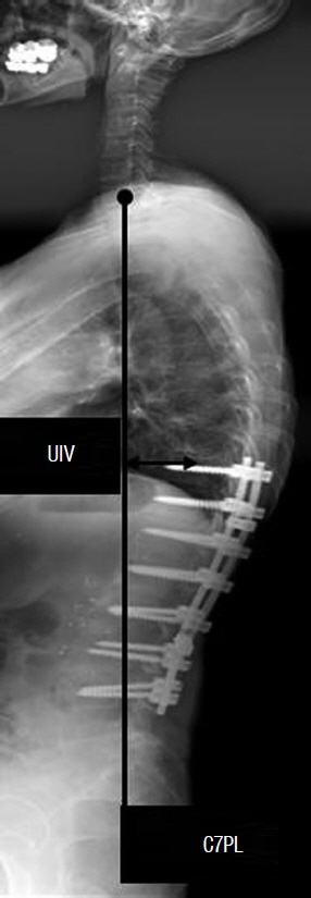
Fig. 1 C7PL was defined distance between C7 plumb line which is the vertical line originating the center of the C7 vertebral body and the posterior superior corner of S1, and its length was measured preoperatively and at ÿnal follow-up. The distance between the center of the UIV and C7PL was also measured early postoperatively and at final follow-up. C7PL: C7 plumb line, UIV: uppermost instrumented vertebra.
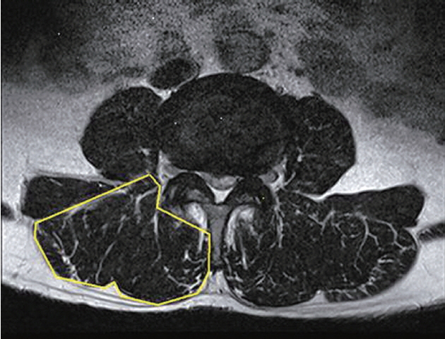
Fig. 2 Thoracolumbar back muscle volumes were measured from T10 to L2 in T2 weighted magnetic resonance images.

Fig. 3 Di°erences in muscle volume (mm2) of two study groups. PJK: proximal junctional kyphosis.

Fig. 4 Scatter plot showed correlation between preoperative C7PL (mm) and mean muscle volume (mm2).
Reference
-
References
1. Beneke R, Neuerburg J, Bohndorf K. Muscle cross-section measurement by magnetic resonance imaging. Eur J Appl Physiol Occup Physiol. 63:424–429. 1991.
Article2. Terracciano C, Celi M, Lecce D, Baldi J, Rastelli E, Lena E, et al. Differential features of muscle fiber atrophy in osteoporosis and osteoarthritis. Osteoporos Int. 24:1095–1100. 2013.
Article3. Cho SK, Shin JI, Kim YJ. Proximal junctional kyphosis following adult spinal deformity surgery. Eur Spine J. 23:2726–2736. 2014.
Article4. Cholewicki J, Panjabi MM, Khachatryan A. Stabilizing function of trunk flexor-extensor muscles around a neutral spine posture. Spine (Phila Pa 1976). 22:2207–2212. 1997.
Article5. Glattes RC, Bridwell KH, Lenke LG, Kim YJ, Rinella A, Edwards C 2nd. Proximal junctional kyphosis in adult spinal deformity following long instrumented posterior spinal fusion: incidence, outcomes, and risk factor analysis. Spine (Phila Pa 1976). 30:1643–1649. 2005.
Article6. Hyun SJ, Rhim SC. Clinical outcomes and complications after pedicle subtraction osteotomy for fixed sagittal imbalance patients: a long-term follow-up data. J Korean Neurosurg Soc. 47:95–101. 2010.
Article7. Harrison DE, Colloca CJ, Harrison DD, Janik TJ, Haas JW, Keller TS. Anterior thoracic posture increases thoracolumbar disc loading. Eur Spine J. 14:234–242. 2005.
Article8. Kang CH, Shin MJ, Kim SM, Lee SH, Lee CS. MRI of paraspinal muscles in lumbar degenerative kyphosis patients and control patients with chronic low back pain. Clin Radiol. 62:479–486. 2007.
Article9. Kim HJ, Bridwell KH, Lenke LG, Park MS, Ahmad A, Song KS, et al. Proximal junctional kyphosis results in inferior SRS pain subscores in adult deformity patients. Spine (Phila Pa 1976). 38:896–901. 2013.
Article10. Kim YJ, Bridwell KH, Lenke LG, Glattes CR, Rhim S, Cheh G. Proximal junctional kyphosis in adult spinal deformity after segmental posterior spinal instrumentation and fusion: minimum five-year follow-up. Spine (Phila Pa 1976). 33:2179–2184. 2008.
Article11. Le Huec JC, Saddiki R, Franke J, Rigal J, Aunoble S. Equilibrium of the human body and the gravity line: the basics. Eur Spine J. 20(Suppl 5):558–563. 2011.
Article12. Lee GA, Betz RR, Clements DH 3rd, Huss GK. Proximal kyphosis after posterior spinal fusion in patients with idiopathic scoliosis. Spine (Phila Pa 1976). 24:795–799. 1999.
Article13. Legaye J, Duval-Beaupère G, Hecquet J, Marty C. Pelvic incidence: a fundamental pelvic parameter for three-dimensional regulation of spinal sagittal curves. Eur Spine J. 7:99–103. 1998.
Article14. Lonner BS, Newton P, Betz R, Scharf C, O’Brien M, Sponseller P, et al. Operative management of Scheuermann’s kyphosis in 78 patients: radiographic outcomes, complications, and technique. Spine (Phila Pa 1976). 32:2644–2652. 2007.15. Maruo K, Ha Y, Inoue S, Samuel S, Okada E, Hu SS, et al. Predictive factors for proximal junctional kyphosis in long fusions to the sacrum in adult spinal deformity. Spine (Phila Pa 1976). 38:E1469–E1476. 2013.
Article16. Roussouly P, Gollogly S, Noseda O, Berthonnaud E, Dimnet J. The vertical projection of the sum of the ground reactive forces of a standing patient is not the same as the C7 plumb line: a radiographic study of the sagittal alignment of 153 asymptomatic volunteers. Spine (Phila Pa 1976). 31:E320–E325. 2006.17. Roussouly P, Pinheiro-Franco JL. Sagittal parameters of the spine: biomechanical approach. Eur Spine J. 20(Suppl 5):578–585. 2011.
Article18. Takemitsu Y, Harada Y, Iwahara T, Miyamoto M, Miyatake Y. Lumbar degenerative kyphosis. Clinical, radiological and epidemiological studies. Spine (Phila Pa 1976). 13:1317–1326. 1988.19. Yagi M, King AB, Boachie-Adjei O. Incidence, risk factors, and natural course of proximal junctional kyphosis: surgical outcomes review of adult idiopathic scoliosis. Minimum 5 years of follow-up. Spine (Phila Pa 1976). 37:1479–1489. 2012.
Article
- Full Text Links
-
- Actions
-
Cited
- CITED
-
- Close
- Share
-
- Similar articles
-
- Proximal Junctional Kyphosis: Diagnosis, Pathogenesis, and Treatment
- Proximal Junctional Kyphosis in Adult Spinal Deformity: Definition, Classification, Risk Factors, and Prevention Strategies
- Proximal Junctional Kyphosis and Proximal Junctional Failure Following Adult Spinal Deformity Surgery
- Risk Factor Analysis of Proximal Junctional Kyphosis after Surgical Treatment of Adult Spinal Deformity with Oblique Lateral Interbody Fusion
- Ligament Augmentation With Mersilene Tape Reduces the Rates of Proximal Junctional Kyphosis and Failure in Adult Spinal Deformity
