J Korean Med Sci.
2016 Jul;31(7):1089-1093. 10.3346/jkms.2016.31.7.1089.
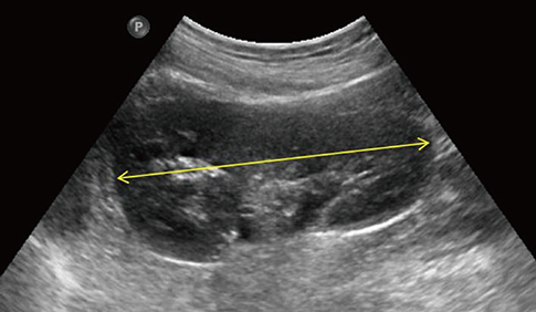
Sonographic Growth Charts for Kidney Length in Normal Korean Children: a Prospective Observational Study
- Affiliations
-
- 1Department of Pediatrics, Jeju National University Hospital, Jeju, Korea.
- 2Department of Internal Medicine, Jeju National University School of Medicine, Jeju, Korea.
- 3Department of Pediatrics, Jeju National University School of Medicine, Jeju, Korea. hansyang78@gmail.com
- 4Department of Diagnostic Radiology, Jeju National University School of Medicine, Jeju, Korea.
- KMID: 2373728
- DOI: http://doi.org/10.3346/jkms.2016.31.7.1089
Abstract
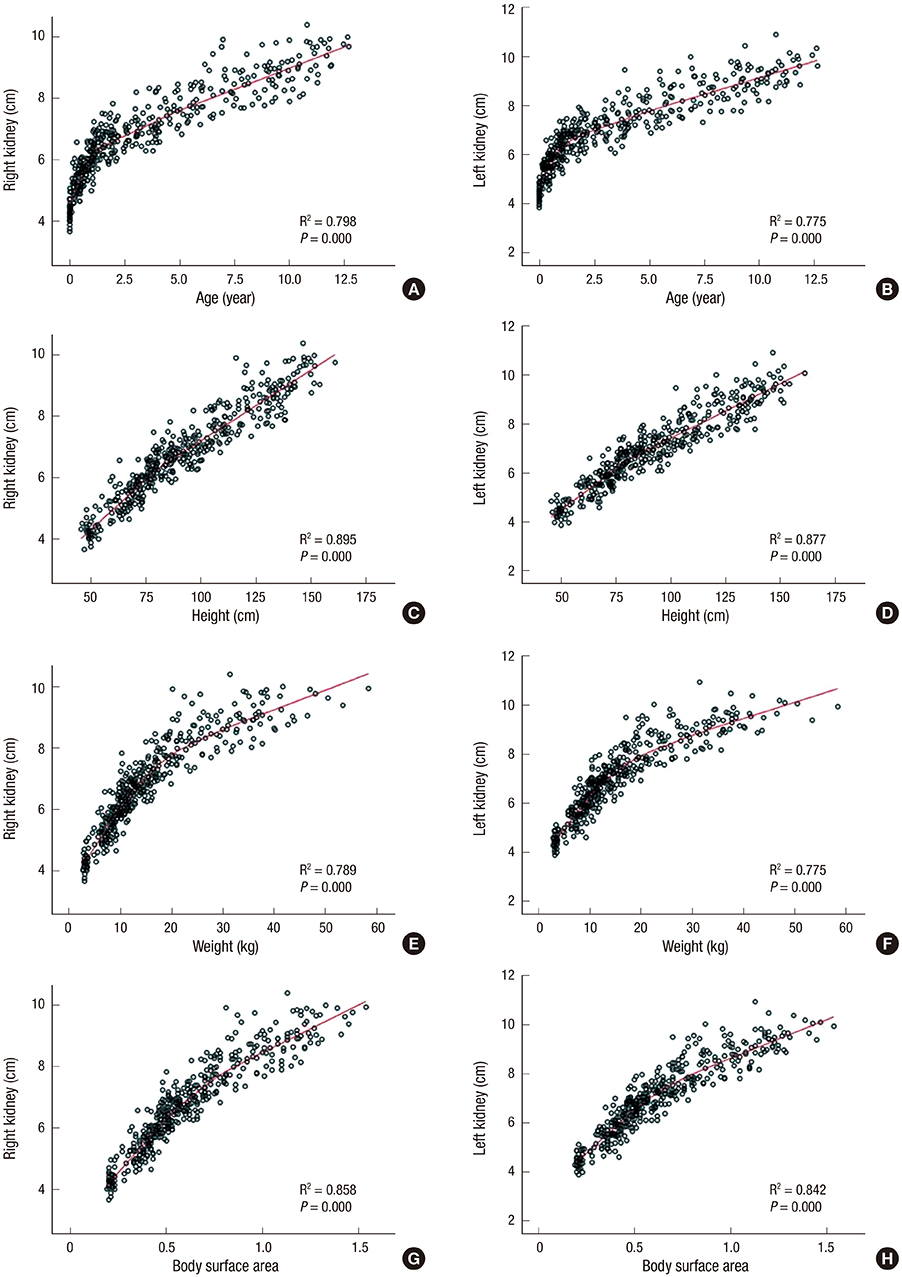
- Kidney length is the most useful parameter for clinical measurement of kidney size, and is useful to distinguish acute kidney injury from chronic kidney disease. In this prospective observational study of 437 normal children aged between 0 and < 13 years, kidney length was measured using sonography. There were good correlations between kidney length and somatic values, including age, weight, height, and body surface area. The rapid growth of height during the first 2 years of life was intimately associated with a similar increase in kidney length, suggesting that height should be considered an important factor correlating with kidney length. Based on our findings, the following regression equation for the reference values of bilateral kidney length for Korean children was obtained: kidney length of the right kidney (cm) = 0.051 × height (cm) + 2.102; kidney length of the left kidney (cm) = 0.051 × height (cm) + 2.280. This equation may aid in the diagnosis of various kidney disorders.
Keyword
MeSH Terms
Figure
Reference
-
1. Queisser-Luft A, Stolz G, Wiesel A, Schlaefer K, Spranger J. Malformations in newborn: results based on 30,940 infants and fetuses from the Mainz congenital birth defect monitoring system (1990-1998). Arch Gynecol Obstet. 2002; 266:163–167.2. Seikaly MG, Ho PL, Emmett L, Fine RN, Tejani A. Chronic renal insufficiency in children: the 2001 Annual Report of the NAPRTCS. Pediatr Nephrol. 2003; 18:796–804.3. Cain JE, Di Giovanni V, Smeeton J, Rosenblum ND. Genetics of renal hypoplasia: insights into the mechanisms controlling nephron endowment. Pediatr Res. 2010; 68:91–98.4. Emamian SA, Nielsen MB, Pedersen JF. Intraobserver and interobserver variations in sonographic measurements of kidney size in adult volunteers. A comparison of linear measurements and volumetric estimates. Acta Radiol. 1995; 36:399–401.5. Schlesinger AE, Hernandez RJ, Zerin JM, Marks TI, Kelsch RC. Interobserver and intraobserver variations in sonographic renal length measurements in children. AJR Am J Roentgenol. 1991; 156:1029–1032.6. Faubel S, Patel NU, Lockhart ME, Cadnapaphornchai MA. Renal relevant radiology: use of ultrasonography in patients with AKI. Clin J Am Soc Nephrol. 2014; 9:382–394.7. Dinkel E, Ertel M, Dittrich M, Peters H, Berres M, Schulte-Wissermann H. Kidney size in childhood. Sonographical growth charts for kidney length and volume. Pediatr Radiol. 1985; 15:38–43.8. Konuş OL, Ozdemir A, Akkaya A, Erbaş G, Celik H, Işik S. Normal liver, spleen, and kidney dimensions in neonates, infants, and children: evaluation with sonography. AJR Am J Roentgenol. 1998; 171:1693–1698.9. Han BK, Babcock DS. Sonographic measurements and appearance of normal kidneys in children. AJR Am J Roentgenol. 1985; 145:611–616.10. Moon JS, Lee SY, Nam CM, Choi JM, Choe BK, Seo JW, Oh K, Jang MJ, Hwang SS, Yoo MH, et al. 2007 Korean National Growth Charts: review of developmental process and an outlook. Korean J Pediatr. 2008; 51:1–25.11. Kim JH, Kim MJ, Lim SH, Kim J, Lee MJ. Length and volume of morphologically normal kidneys in Korean children: ultrasound measurement and estimation using body size. Korean J Radiol. 2013; 14:677–682.12. Zerin JM, Blane CE. Sonographic assessment of renal length in children: a reappraisal. Pediatr Radiol. 1994; 24:101–106.13. Kim IO, Cheon JE, Lee YS, Lee SW, Kim OH, Kim JH, Kim HD, Sim JS. Kidney length in normal Korean children. J Korean Soc Ultrasound Med. 2010; 29:181–188.14. Lee MJ, Son MK, Kwak BO, Park HW, Chung S, Kim KS. Kidney size estimation in Korean children with Technesium-99m dimercaptosuccinic acid scintigraphy. Korean J Pediatr. 2014; 57:41–45.15. Mesrobian HG, Laud PW, Todd E, Gregg DC. The normal kidney growth rate during year 1 of life is variable and age dependent. J Urol. 1998; 160:989–993.16. Akhavan A, Brajtbord JS, McLeod DJ, Kabarriti AE, Rosenberg HK, Stock JA. Simple, age-based formula for predicting renal length in children. Urology. 2011; 78:405–410.17. Haugstvedt S, Lundberg J. Kidney size in normal children measured by sonography. Scand J Urol Nephrol. 1980; 14:251–255.18. Ryoo NY, Shin HY, Kim JH, Moon JS, Lee CG. Change in the height of Korean children and adolescents: analysis from the Korea National Health and Nutrition Survey II and V. Korean J Pediatr. 2015; 58:336–340.19. Otiv A, Mehta K, Ali U, Nadkarni M. Sonographic measurement of renal size in normal Indian children. Indian Pediatr. 2012; 49:533–536.
- Full Text Links
- Actions
-
Cited
- CITED
-
- Close
- Share
- Similar articles
-
- Reappraisal of Regional Growth Charts in the Era of WHO Growth Standards
- Monitoring Growth in Childhood: Practical Clinical Guide
- Roentgenographic Measurement of Normal Kidney Size in Korean Children
- The radiographic estimation of the kidney in normal Korean children
- Development of disease-specific growth charts in Turner syndrome and Noonan syndrome


