Development and Evaluation of a Duplex Real-Time PCR Assay With a Novel Internal Standard for Precise Quantification of Plasma DNA
- Affiliations
-
- 1Department of Laboratory Medicine, the First Affiliated Hospital of Nanjing Medical University, Nanjing, China. sypan@njmu.edu.cn
- 2National Key Clinical Department of Laboratory Medicine, the First Affiliated Hospital of Nanjing Medical University, Nanjing, China.
- KMID: 2373609
- DOI: http://doi.org/10.3343/alm.2017.37.1.18
Abstract
- BACKGROUND
Circulating levels of cell-free DNA increase in many pathologic conditions. However, notable discrepancies in the quantitative analysis of cell-free DNA from a large number of laboratories have become a considerable pitfall, hampering its clinical application.
METHODS
We designed a novel recombinant DNA fragment that could be applied as an internal standard in a newly developed and validated duplex real-time PCR assay for the quantitative analysis of total cell-free plasma DNA, which was tested in 5,442 healthy adults and 200 trauma patients.
RESULTS
Compared with two traditional methods, this novel assay showed a lower detection limit of 0.1 ng/mL, lower intra- and inter-assay CVs, and higher accuracy in the recovery test. The median plasma DNA concentration of healthy males (20.3 ng/mL, n=3,092) was significantly higher than that of healthy females (16.1 ng/mL, n=2,350) (Mann-Whitney two-sample rank sum test, P<0.0001). The reference intervals of plasma DNA concentration were 0-45.8 ng/mL and 0-52.5 ng/mL for healthy females and males, respectively. The plasma DNA concentrations of the majority of trauma patients (96%) were higher than the upper normal cutoff values and were closely related to the corresponding injury severity scores (R²=0.916, P<0.0001).
CONCLUSIONS
This duplex real-time PCR assay with a new internal standard could eliminate variation and allow for more sensitive, repeatable, accurate, and stable quantitative measurements of plasma DNA, showing promising application in clinical diagnosis.
MeSH Terms
Figure
-
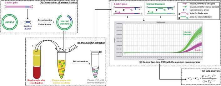
Fig. 1 The schematic concepts of duplex real-time PCR with internal standard for quantitative detection of plasma DNA. The whole process includes 4 steps: (A) A 41-bp artificial double-stranded DNA sequence corresponding to the human β–actin gene is cloned and inserted to pMD18-T vector, which is then linearized by restriction enzyme digestion. (B) The recombinant plasmid DNA is added into the cell-free plasma sample with known concentration and extracted together with endogenous nucleic acids. (C) The internal standard and target gene are then amplified simultaneously in the same tube with the common reverse primer focusing on the 41-bp corresponding sequence by duplex real-time PCR, where fluorescence signals are detected separately (Applied Biosystems 7500 Sequence Detector). (D) The plasma DNA concentrations are calculated according to the internal standard by the equation.
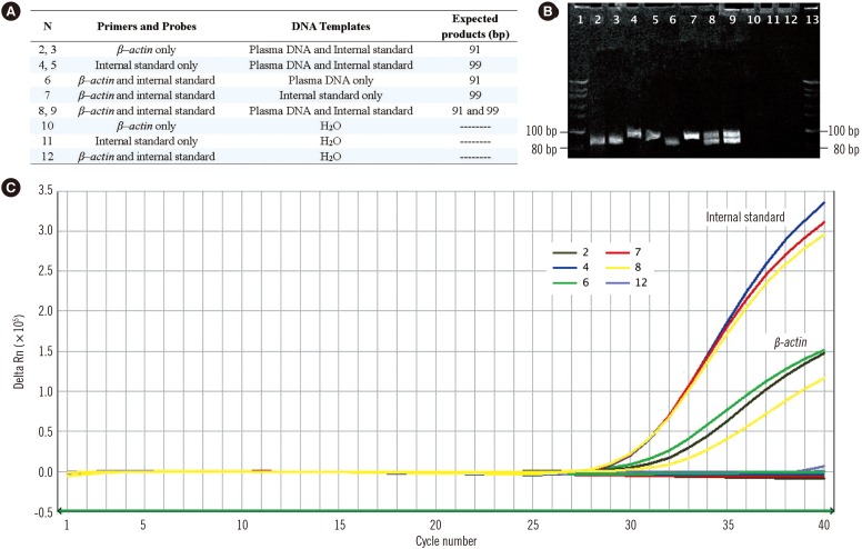
Fig. 2 Evaluation of the specificity of the duplex real-time PCR assay with internal standard. Real-time PCR amplifications of plasma DNA, internal standard DNA, and the mixture of both in a parallel PCR reaction with their corresponding primers and probes are performed in preliminary experiments to confirm the specificity of this novel duplex PCR assay (A). The target β–actin gene and internal standard are amplified by their corresponding primers for 91-bp and 99-bp PCR products respectively (B) and Taqman probes for fluorescent signals released from JOE and FAM reports and detected in channels 2 and 1 of the ABI 7500 Sequence Detector (C).
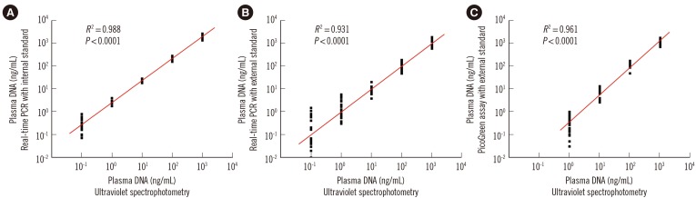
Fig. 3 Evaluation of the sensitivity of the three different plasma DNA quantitative assays. The statistically significant correlations of DNA levels of 10-fold serial diluted plasma samples (X-axis, common logarithmic scale) with quantitative results (Y-axis, common logarithmic scale) determined by the novel duplex real-time PCR assay with (A) internal standard, (B) the single real-time PCR assay with external standards, and (C) the dye PicoGreen assay.
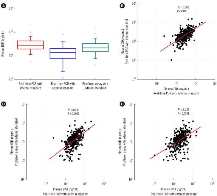
Fig. 4 Quantitative analysis of plasma DNA in the plasma of 400 healthy adults by three methods. While the median plasma DNA concentration of 400 healthy adults determined by duplex real time PCR with internal standard (18.1 ng/mL) was significantly higher than those determined by real time PCR and PicoGreen assay with external standards (8.2 ng/ml and 13.3 ng/ml; Wilcoxon matched-pairs signed-rank test, P=0.0000) (A), there were statistically significant but weak positive correlations among plasma DNA levels determined by these three different methods (B-D).
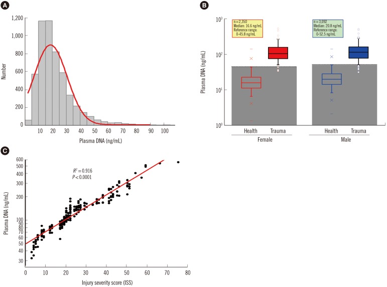
Fig. 5 Quantitative analysis of plasma DNA in 5,443 healthy adults and 200 trauma patients by the duplex real time PCR assay with internal standard. (A) The histogram shows a positively skewed distribution of populations in the different levels of plasma DNA. (B) Box-plots of plasma DNA in healthy adults and trauma patients with different genders shows the reference intervals of plasma DNA concentration are 0-45.8 ng/mL and 0-52.5 ng/mL for healthy females and males, respectively, and the plasma DNA concentrations of most trauma patients (96%) are higher than the upper reference ranges. (C) A statistically significant positive correlation is revealed between plasma DNA concentrations in trauma patients with their ISS values (Spearman rank correlation test, R2=0.916, P<0.0001).
Reference
-
1. Mandel P, Metais P. Nucleic acids in plasma samples from human blood. C R Seances Soc Biol Fil. 1948; 142:241–243. PMID: 18875018.2. Leon SA, Shapiro B, Sklaroff DM, Yaros MJ. Free DNA in the serum of cancer patients and the effect of therapy. Cancer Res. 1977; 37:646–650. PMID: 837366.3. Tong YK, Lo YM. Diagnostic developments involving cell-free (circulating) nucleic acids. Clin Chim Acta. 2006; 363:187–196. PMID: 16126188.4. Tissot C, Toffart AC, Villar S, Souquet PJ, Merle P, Moro-Sibilot D, et al. Circulating free DNA concentration is an independent prognostic biomarker in lung cancer. Eur Respir J. 2015; 46:1773–1780. PMID: 26493785.5. Sozzi G, Roz L, Conte D, Mariani L, Andriani F, Lo Vullo S, et al. Plasma DNA quantification in lung cancer computed tomography screening: five-year results of a prospective study. Am J Respir Crit Care Med. 2009; 179:69–74. PMID: 18787214.6. Altimari A, Grigioni AD, Benedettini E, Gabusi E, Schiavina R, Martinelli A, et al. Diagnostic role of circulating free plasma DNA detection in patients with localized prostate cancer. Am J Clin Pathol. 2008; 129:756–762. PMID: 18426736.7. Szpechcinski A, Chorostowska-Wynimko J, Struniawski R, Kupis W, Rudzinski P, Langfort R, et al. Cell-free DNA levels in plasma of patients with non-small-cell lung cancer and inflammatory lung disease. Br J Cancer. 2015; 113:476–483. PMID: 26125447.8. Paci M, Maramotti S, Bellesia E, Formisano D, Albertazzi L, Ricchetti T, et al. Circulating plasma DNA as diagnostic biomarker in non-small cell lung cancer. Lung Cancer. 2009; 64:92–97. PMID: 18804892.9. Gautschi O, Bigosch C, Huegli B, Jermann M, Marx A, Chassé E, et al. Circulating deoxyribonucleic acid as prognostic marker in non-small-cell lung cancer patients undergoing chemotherapy. J Clin Oncol. 2004; 22:4157–4164. PMID: 15483026.10. Skvortsova TE, Rykova EY, Tamkovich SN, Bryzgunova OE, Starikov AV, Kuznetsova NP, et al. Cell-free and cell-bound circulating DNA in breast tumours: DNA quantification and analysis of tumour-related gene methylation. Br J Cancer. 2006; 94:1492–1495. PMID: 16641902.11. Pinzani P, Salvianti F, Zaccara S, Massi D, De Giorgi V, Pazzagli M, et al. Circulating cell-free DNA in plasma of melanoma patients: qualitative and quantitative considerations. Clin Chim Acta. 2011; 412:2141–2145. PMID: 21839068.12. Lo YM, Tein MS, Lau TK, Haines CJ, Leung TN, Poon PM, et al. Quantitative analysis of fetal DNA in maternal plasma and serum: implications for noninvasive prenatal diagnosis. Am J Hum Genet. 1998; 62:768–775. PMID: 9529358.13. Manokhina I, Singh TK, Penaherrera MS, Robinson WP. Quantification of cell-free DNA in normal and complicated pregnancies: overcoming biological and technical issues. PLoS One. 2014; 9:e101500. PMID: 24987984.14. Lo YM, Rainer TH, Chan LY, Hjelm NM, Cocks RA. Plasma DNA as a prognostic marker in trauma patients. Clin Chem. 2000; 46:319–323. PMID: 10702517.15. Rainer TH. Plasma DNA, prediction and post-traumatic complications. Clin Chim Acta. 2001; 313:81–85. PMID: 11694243.16. Saukkonen K, Lakkisto P, Varpula M, Varpula T, Voipio-Pulkki LM, Pettilä V, et al. Association of cell-free plasma DNA with hospital mortality and organ dysfunction in intensive care unit patients. Intensive Care Med. 2007; 33:1624–1627. PMID: 17541553.17. Chen JA, Meister S, Urbonaviciute V, Rodel F, Wilhelm S, Kalden JR, et al. Sensitive detection of plasma/serum DNA in patients with systemic lupus erythematosus. Autoimmunity. 2007; 40:307–310. PMID: 17516216.18. Macher HC, Suárez-Artacho G, Guerrero JM, Gomez-Bravo MA, Alvarez-Gómez S, Bernal-Bellido C, et al. Monitoring of transplanted liver health by quantification of organ-specific genomic marker in circulating DNA from receptor. PLoS One. 2014; 9:e113987. PMID: 25489845.19. Emons H, Corbisier P. Nucleic acid quantification-progress and pitfalls. Anal Bioanal Chem. 2014; 406:6469–6470. PMID: 25148828.20. Gormally E, Hainaut P, Caboux E, Airoldi L, Autrup H, Malaveille C, et al. Amount of DNA in plasma and cancer risk: a prospective study. Int J Cancer. 2004; 111:746–749. PMID: 15252845.21. de Kok JB, Hendriks JC, van Solinge WW, Willems HL, Mensink EJ, Swinkels DW. Use of real-time quantitative PCR to compare DNA isolation methods. Clin Chem. 1998; 44:2201–2204. PMID: 9761259.22. Jorgez CJ, Dang DD, Simpson JL, Lewis DE, Bischoff FZ. Quantity versus quality: optimal methods for cell-free DNA isolation from plasma of pregnant women. Genet Med. 2006; 8:615–619. PMID: 17079877.23. Szpechcinski A, Struniawska R, Zaleska J, Chabowski M, Orlowski T, Roszkowski K, et al. Evaluation of fluorescence-based methods for total vs. amplifiable DNA quantification in plasma of lung cancer patients. J Physiol Pharmacol. 2008; 59(S6):675–681. PMID: 19218694.24. Holdenrieder S, Stieber P, Chan LY, Geiger S, Kremer A, Nagel D, et al. Cell-free DNA in serum and plasma: comparison of ELISA and quantitative PCR. Clin Chem. 2005; 51:1544–1546. PMID: 16040855.25. Mauger F, Dulary C, Daviaud C, Deleuze JF, Tost J. Comprehensive evaluation of methods to isolate, quantify, and characterize circulating cell-free DNA from small volumes of plasma. Anal Bioanal Chem. 2015; 407:6873–6878. PMID: 26123439.26. Devonshire AS, Whale AS, Gutteridge A, Jones G, Cowen S, Foy CA, et al. Towards standardisation of cell-free DNA measurement in plasma: controls for extraction efficiency, fragment size bias and quantification. Anal Bioanal Chem. 2014; 406:6499–6512. PMID: 24853859.27. Ramakers C, Ruijter JM, Deprez RH, Moorman AF. Assumption-free analysis of quantitative real-time polymerase chain reaction (PCR) data. Neurosci Lett. 2003; 339:62–66. PMID: 12618301.28. Xue X, Teare MD, Holen I, Zhu YM, Woll PJ. Optimizing the yield and utility of circulating cell-free DNA from plasma and serum. Clin Chim Acta. 2009; 404:100–104. PMID: 19281804.29. Zhang P, Ren J, Shen Z. A new quantitative method for circulating DNA level in human serum by capillary zone electrophoresis with laser-induced fluorescence detection. Electrophoresis. 2004; 25:1823–1828. PMID: 15213980.30. Fujii S, Inagaki K, Miyashita S, Nagasawa K, Chiba K, Takatsu A. A coupling system of capillary gel electrophoresis with inductively coupled plasma-mass spectrometry for the determination of double stranded DNA fragments. Metallomics. 2013; 5:424–428. PMID: 23604270.31. Camp CL, Sharp BL, Reid HJ, Entwisle J, Goenaga-Infante H. Analysis of mono-phosphate nucleotides as a potential method for quantification of DNA using high performance liquid chromatography-inductively coupled plasma-mass spectrometry. Anal Bioanal Chem. 2012; 402:367–372. PMID: 21877184.32. Huang CZ, Li KA, Tong SY. Determination of nucleic acids by a resonance light-scattering technique with alpha, beta, gamma, delta-tetrakis [4-(trimethylammoniumyl)phenyl]porphine. Anal Chem. 1996; 68:2259–2263. PMID: 9027231.33. Liu R, Yang J, Wu X, Sun C. Study on the resonance light scattering spectrum of berberine-cetyltrimethylammonium bromide system and the determination of nucleic acids at nanogram levels. Spectrochim Acta A Mol Biomol Spectrosc. 2002; 58:457–465. PMID: 11905531.34. Watts HJ, Yeung D, Parkes H. Real-time detection and quantification of DNA hybridization by an optical biosensor. Anal Chem. 1995; 67:4283–4289. PMID: 8633773.35. Pallisgaard N, Spindler KL, Andersen RF, Brandslund I, Jakobsen A. Controls to validate plasma samples for cell free DNA quantification. Clin Chim Acta. 2015; 446:141–146. PMID: 25896958.36. Wu TL, Zhang D, Chia JH, Tsao K, Sun CF, Wu JT. Cell-free DNA: measurement in various carcinoma and establishment of normal reference range. Clin Chim Acta. 2002; 321:77–87. PMID: 12031596.37. Lam NY, Rainer TH, Chan LY, Joynt GM, Lo YM. Time course of early and late changes in plasma DNA in trauma patients. Clin Chem. 2003; 49:1286–1291. PMID: 12881444.
- Full Text Links
-
- Actions
-
Cited
- CITED
-
- Close
- Share
-
- Similar articles
-
- Influence of Standard Curves on Relative Quantification using Real-time PCR
- Comparison between Real-Time PCR and Agarose Gel Electrophoresis for DNA Quantification
- Performance Evaluation of the Real-Q Cytomegalovirus (CMV) Quantification Kit Using Two Real-Time PCR Systems for Quantifying CMV DNA in Whole Blood
- Performance Evaluation of Real-Q HBV Quantification Kit for HBV DNA by Real-Time PCR
- Performance of the Real-Q EBV Quantification Kit for Epstein-Barr Virus DNA Quantification in Whole Blood
