J Gastric Cancer.
2012 Mar;12(1):43-45.
Mesenteric Pseudocyst of the Small Bowel in Gastric Cancer Patient: A Case Report
- Affiliations
-
- 1Department of Surgery, Konyang University College of Medicine, Daejeon, Korea. cwj@kyuh.ac.kr
- 2Division of Gastroenterology and Hepatology, Konyang University College of Medicine, Daejeon, Korea.
- 3Depatrment of Pathology, Konyang University College of Medicine, Daejeon, Korea.
- 4Sokpyunhan Internal Medicine Clinic, Daejeon, Korea.
Abstract
- Mesenteric pseudocyst is rare. This term is used to describe the abdominal cystic mass, without the origin of abdominal organ. We presented a case of mesenteric pseudocyst of the small bowel in a 70-year-old man. Esophago-gastro-duodenoscopy showed a 3.5 cm sized excavated lesion on the posterior wall of angle. Endocopic biopsy confirmed a histologic diagnosis of the poorly differentiated adenocarcinoma, which includes the signet ring cell component. Abdominal computed tomography scan showed a focal mucosal enhancement in the posterior wall of angle of the stomach, a 2.4 cm sized enhancing mass on the distal small bowel loop, without distant metastases or ascites in rectal shelf, and multiple gallbladder stones. The patient underwent subtotal gastrectomy with gastroduodenostomy, segmental resection of the small bowel, and cholecystectomy. The final pathological diagnosis was mesenteric pseudocyst. This is the first case report describing incidentally detected mesenteric pseudocyst of the small bowel in gastric cancer patients.
Keyword
MeSH Terms
Figure
-

Fig. 1 Preoperative computed tomography.
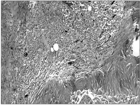
Fig. 2 Fibrous cystic wall without specific endothelial or epithelial lining epithelium. Cystic content shows cholesterol cleft (H&E, ×12.5).
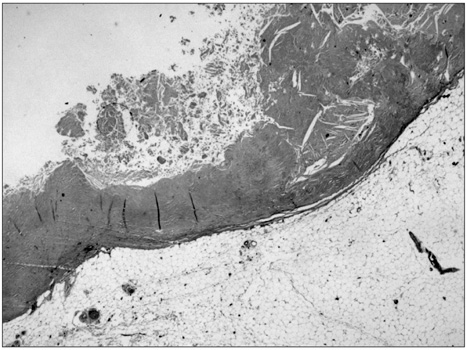
Fig. 3 Fibrous cystic wall and focal foam cell collection (H&E, ×40).
Reference
-
1. Gallego JC, González JM, Fernández-Virgós A, del Castillo M. Retrorectal mesenteric cyst (non-pancreatic pseudocyst) in adult. Eur J Radiol. 1996. 23:135–137.
Article2. Ros PR, Olmsted WW, Moser RP Jr, Dachman AH, Hjermstad BH, Sobin LH. Mesenteric and omental cysts: histologic classification with imaging correlation. Radiology. 1987. 164:327–332.
Article3. Kurtz RJ, Heimann TM, Holt J, Beck AR. Mesenteric and retroperitoneal cysts. Ann Surg. 1986. 203:109–112.
Article4. Iida T, Suenaga M, Takeuchi Y, Kobayashi T, Tobinaga J, Sakai M, et al. Mesenteric pseudocyst of the sigmoid colon. J Gastroenterol. 2003. 38:1081–1085.
Article5. Fan HL, Chen TW, Hong ZJ, Hsieh CB, Chan DC, Chen CJ, et al. Volvulus of small intestine: rare complication of mesenteric pseudocyst. Z Gastroenterol. 2009. 47:1208–1210.
Article
- Full Text Links
-
- Actions
-
Cited
- CITED
-
- Close
- Share
-
- Similar articles
-
- Mesenteric and Omental Cyst: CT Findings
- An Unusual Cause of Mesenteric Ischemia of the Small Intestine: Jejunal Neuroendocrine Tumor
- Delayed Small Bowel Ischemia following Minor Mesenteric Injury
- Cytomegalovirus Enteritis in an Immunocompetent Patient Causing Small Bowel Obstruction and Superior Mesenteric Artery Thrombosis: A Case Report
- A Case of Small Bowel GIST Initially Suspected as Peritoneal Seeding of Gastric Cancer
