Inflammatory Myofibroblastic Tumor Treated with Laparoscopic Proximal Gastrectomy and Double-Tract Anastomosis
- Affiliations
-
- 1Department of Surgery, Yeouido St. Mary's Hospital, College of Medicine, The Catholic University of Korea, Seoul, Korea. kimwook@catholic.ac.kr
- KMID: 2372373
- DOI: http://doi.org/10.5230/jgc.2015.15.1.64
Abstract
- Inflammatory myofibroblastic tumors (IMTs) of the stomach are extremely rare in adults, and their oncologic prognosis is not well understood. We present a 28-year-old man with a proximal gastric IMT. The patient visited the emergency department of Yeouido St. Mary's Hospital with syncope and hematemesis. Hemoglobin levels were <5.5 g/dl. Gastric fibroscopy showed a protruding mass 4x4 cm in size, with central ulceration on the posterior wall of the fundus and diffuse wall thickening throughout the cardia and anterior wall of the upper body. Endoscopic biopsy revealed indeterminate spindle cells, along with inflammation. Given the risk of rebleeding, an operation was performed despite the uncertain diagnosis. Because the mass was circumferential, laparoscopic proximal gastrectomy and double-tract anastomosis were performed to ensure a safe resection margin. The pathological diagnosis was consistent with an IMT originating from the stomach, although the tumor was negative for anaplastic lymphoma kinase gene mutation.
Keyword
MeSH Terms
Figure
-
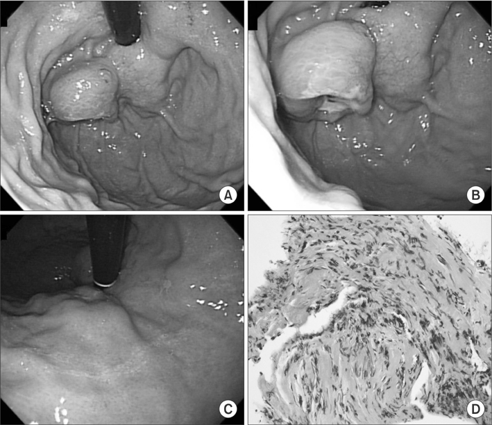
Fig. 1 Preoperative endoscopic and biopsy findings. (A, B) Protruding mass 4×4 cm in size with central ulceration. (C) Two submucosal tumors on the lesser curvature side of the cardia and upper body. (D) Histological examination of the fundic mass revealed nonspecific spindle cells and inflammatory cells (H&E, ×100).
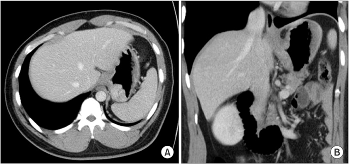
Fig. 2 Computed tomography scan shows fundic mass with diffusely thickened gastric wall on the lesser curvature side of the upper body and cardia. (A) Axial image. (B) Coronal image.
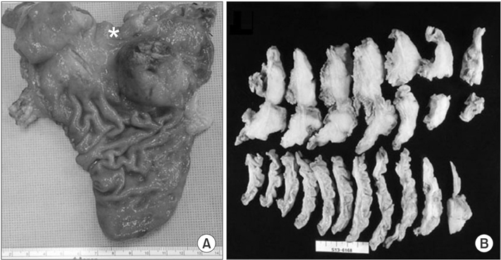
Fig. 3 (A) Opened specimen shows protruding mass on posterior wall of fundus and diffuse wall thickening of cardia and high body (*esophageal mucosa). (B) Cut section shows large mass with infiltration into the muscle layer.

Fig. 4 Histological findings show spindle-shaped tumor cells under a myxoid stroma with lymphoplasmacytic infiltration among the tumor cells. (A) H&E, ×40. (B) H&E, ×100. (C) H&E, ×400. (D) Actin (+), ×400. (E) CD117 (-), ×400. (F) Anaplastic lymphoma kinase mutation (-), ×400. (G) IgG (+), ×400. (H) IgG4 (+), ×400; IgG4/IgG ratio <0.4. IgG = immunoglobulin G.
Reference
-
1. Shi H, Wei L, Sun L, Guo A. Primary gastric inflammatory myofibroblastic tumor: a clinicopathologic and immunohistochemical study of 5 cases. Pathol Res Pract. 2010; 206:287–291.2. Lee WA, Lee MK, Jeen YM, Kie JH, Chung JJ, Yun SH. Solitary fibrous tumor arising in gastric serosa. Pathol Int. 2004; 54:436–439.3. Coffin CM, Hornick JL, Fletcher CD. Inflammatory myofibroblastic tumor: comparison of clinicopathologic, histologic, and immunohistochemical features including ALK expression in atypical and aggressive cases. Am J Surg Pathol. 2007; 31:509–520.4. Kim EY, Lee IK, Lee YS, Yang N, Chung DJ, Yim KI, et al. Inflammatory myofibroblastic tumor in colon. J Korean Surg Soc. 2012; 82:45–49.5. Qiu JF, Shi YJ, Fang L, Wang HF, Zhang MC. High fever as an initial symptom of primary gastric inflammatory myofibroblastic tumor in an adult woman. Int J Clin Exp Med. 2014; 7:1468–1473.6. Katakwar A, Gedam BS, Mukewar S, Agasti A. Primary gastric inflammatory myofibroblastic tumor in an adult-case report with brief review. Indian J Surg Oncol. 2014; 5:66–70.7. Cook JR, Dehner LP, Collins MH, Ma Z, Morris SW, Coffin CM, et al. Anaplastic lymphoma kinase (ALK) expression in the inflammatory myofibroblastic tumor: a comparative immunohistochemical study. Am J Surg Pathol. 2001; 25:1364–1371.8. Jain A, Kasana S, Ramrakhiani D, Sharma M. Inflammatory myofibroblastic tumor of the stomach in an adult female: report of a rare case and review of the literature. Turk J Gastroenterol. 2012; 23:399–405.9. Park SH, Kim JH, Min BW, Song TJ, Son GS, Kim SJ, et al. Exophytic inflammatory myofibroblastic tumor of the stomach in an adult woman: a rare cause of hemoperitoneum. World J Gastroenterol. 2008; 14:136–139.10. Coffin CM, Watterson J, Priest JR, Dehner LP. Extrapulmonary inflammatory myofibroblastic tumor (inflammatory pseudotumor). A clinicopathologic and immunohistochemical study of 84 cases. Am J Surg Pathol. 1995; 19:859–872.11. Coffin CM, Fletcher JA. Inflammatory myofibroblastic tumour. In : Fletcher CDM, Unni KK, Mertens F, editors. World Health Organization. International Agency for Research on Cancer. Pathology and Genetics of Tumours of Soft Tissue and Bone. Lyon: IARC Press;2002. p. 91–93.
- Full Text Links
-
- Actions
-
Cited
- CITED
-
- Close
- Share
-
- Similar articles
-
- Single-Port Laparoscopic Proximal Gastrectomy with Double Tract Reconstruction for Early Gastric Cancer: Report of a Case
- Laparoscopic Proximal Gastrectomy as a Surgical Treatment for Upper Third Early Gastric Cancer
- Case Report: Gastrobronchial Fistula after Sleeve Gastrectomy: Treated by Laparoscopic Proximal Gastrectomy with Double Tract Reconstruction
- Intracorporeal Anastomosis in Laparoscopic Gastric Cancer Surgery
- Inflammatory Myofibroblastic Tumor of Nasal Septum after Septoplasty: A Case Report
