Ann Lab Med.
2015 Sep;35(5):500-505. 10.3343/alm.2015.35.5.500.
Evaluation of Dual-Color Fluorescence In Situ Hybridization With Peptide Nucleic Acid Probes for the Detection of Mycobacterium tuberculosis and Non-Tuberculous Mycobacteria in Clinical Specimens
- Affiliations
-
- 1Department of Laboratory Medicine, Pusan National University Yangsan Hospital, Yangsan, Korea. cchl@pusan.ac.kr
- 2Department of Laboratory Medicine, Cheukchu Hospital, Changwon, Korea.
- 3Department of Laboratory Medicine, Pusan National University Hospital, Busan, Korea.
- 4Research Institute for Convergence of Biomedical Science and Technology, Pusan National University Yangsan Hospital, Yangsan, Korea.
- KMID: 2369765
- DOI: http://doi.org/10.3343/alm.2015.35.5.500
Abstract
- BACKGROUND
Peptide nucleic acid (PNA) probes are artificial DNA analogues with a hydrophobic nature that can penetrate the mycobacterial cell wall. We evaluated a FISH method for simultaneous detection and identification of Mycobacterium tuberculosis (MTB) and non-tuberculous mycobacteria (NTM) in clinical respiratory specimens using differentially labeled PNA probes.
METHODS
PNA probes targeting the mycobacterial 16S ribosomal RNA were synthesized. The cross-reactivity of MTB- and NTM-specific probes was examined with reference strains and 10 other frequently isolated bacterial species. A total of 140 sputum specimens were analyzed, comprising 100 MTB-positive specimens, 21 NTM-positive specimens, and 19 MTB/NTM-negative specimens; all of them were previously confirmed by PCR and culture. The PNA FISH test results were graded by using the United States Centers for Disease Control and Prevention-recommended scale and compared with the results from the fluorochrome acid-fast bacterial stain.
RESULTS
The MTB- and NTM-specific PNA probes showed no cross-reactivity with other tested bacterial species. The test results demonstrated 82.9% agreement with the culture results with diagnostic sensitivity of 80.2% and diagnostic specificity of 100.0% (kappa=0.52, 95% confidence interval: 0.370-0.676).
CONCLUSIONS
Dual-color PNA FISH showed high specificity for detecting and identifying mycobacteria in clinical specimens. However, because of its relatively low sensitivity, this method could be more applicable to culture confirmation. In application to direct specimens, the possibility of false-negative results needs to be considered.
Keyword
MeSH Terms
Figure
-
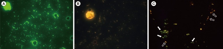
Fig. 1 Peptide nucleic acid (PNA) FISH analysis. (A) Positive PNA FISH for Mycobacterium tuberculosis (MTB). (B) Positive PNA FISH for non-tuberculous mycobacteria (NTM). (C) PNA FISH for a mixture of the MTB ATCC 13950 and M. kansasii ATCC 12479 strains. The MTB PNA probe was labeled with 6-carboxyfluorescein fluorescent dye (green, filled arrows), and the NTM PNA probe was labeled with cyanine 3 fluorescent dye (orange, non-filled arrows).
Reference
-
1. World Health Organization. Global tuberculosis report 2014. Geneva: World Health Organization;2014.2. Shinnick TM, Iademarco MF, Ridderhof JC. National plan for reliable tuberculosis laboratory services using a systems approach. Recommendations from CDC and the Association of Public Health Laboratories Task Force on Tuberculosis Laboratory Services. MMWR Recomm Rep. 2005; 54:1–12. PMID: 15829862.3. Fitzgerald DW, Sterling TR, Hass DW. Mycobacterium tuberculosis. In : Mandell GL, Bennett JE, Dolan R, editors. Mandell, Douglas, and Bennett's principles and practice of infectious diseases. 7th ed. Philadelphia, PA: Elsevier;2010.4. Padilla E, Manterola JM, Rasmussen OF, Lonca J, Domínguez J, Matas L, et al. Evaluation of a fluorescence hybridisation assay using peptide nucleic acid probes for identification and differentiation of tuberculous and non-tuberculous mycobacteria in liquid cultures. Eur J Clin Microbiol Infect Dis. 2000; 19:140–145. PMID: 10746504.
Article5. Perry-O'Keefe H, Rigby S, Oliveira K, Sørensen D, Stender H, Coull J, et al. Identification of indicator microorganisms using a standardized PNA FISH method. J Microbiol Methods. 2001; 47:281–292. PMID: 11714518.6. Stender H, Lund K, Petersen KH, Rasmussen OF, Hongmanee P, Miörner H, et al. Fluorescence In situ hybridization assay using peptide nucleic acid probes for differentiation between tuberculous and nontuberculous mycobacterium species in smears of mycobacterium cultures. J Clin Microbiol. 1999; 37:2760–2765. PMID: 10449448.
Article7. Porcheddu A, Giacomelli G. Peptide nucleic acids (PNAs), a chemical overview. Curr Med Chem. 2005; 12:2561–2599. PMID: 16248816.
Article8. Altschul SF, Gish W, Miller W, Myers EW, Lipman DJ. Basic local alignment search tool. J Mol Biol. 1990; 215:403–410. PMID: 2231712.
Article9. Diagnostic Standards and Classification of Tuberculosis in Adults and Children. This official statement of the American Thoracic Society and the Centers for Disease Control and Prevention was adopted by the ATS Board of Directors, July 1999. This statement was endorsed by the Council of the Infectious Disease Society of America, September 1999. Am J Respir Crit Care Med. 2000; 161:1376–1395. PMID: 10764337.
- Full Text Links
-
- Actions
-
Cited
- CITED
-
- Close
- Share
-
- Similar articles
-
- Evaluation of Peptide Nucleic Acid Probe-Based Fluorescence In Situ Hybridization for the Detection of Mycobacterium tuberculosis Complex and Nontuberculous Mycobacteria in Clinical Respiratory Specimens
- Detection of Mycobactrium tuberculosis by in situ hybridization
- Evaluation of Peptide Nucleic Acid Probe-based Real-time PCR for Detection of Mycobacterium tuberculosis Complex and Nontuberculous Mycobacteria in Respiratory Specimens
- Recent Advances in Tuberculosis and Nontuberculous Mycobacteria Lung Disease
- Comparison of the Rate of Detection of Immunoglobulin Heavy Chain Gene Rearrangement by Fluoresecence In Situ Hybridization Probes in Multiple Myeloma
