Is Carotid Artery Ultrasound Still Useful Method for Evaluation of Atherosclerosis?
- Affiliations
-
- 1Division of Cardiology, Department of Internal Medicine, St. Vincent's Hospital, Suwon, Korea.
- 2Division of Cardiology, Department of Internal Medicine, Seoul St. Mary's Hospital, College of Medicine, The Catholic University of Korea, Seoul, Korea. younhj@catholic.ac.kr
- KMID: 2365273
- DOI: http://doi.org/10.4070/kcj.2016.0232
Abstract
- Carotid ultrasound is an imaging modality that allows non-invasive assessment of vascular anatomy and function. Carotid intima-media thickness (IMT) has been shown to predict cardiovascular (CV) risk in multiple large studies. However, in 2013, American College of Cardiology/American Heart Association guidelines designated that the carotid IMT as class III evidence level was not recommended for use in clinical practice as a routine measurement of risk assessment for a first atherosclerotic CV event. Following the announcement of this guideline, combined common carotid IMT and plaque, including plaque tissue characterization and plaque burden, using 3D ultrasound was reported to be better than either measurement alone in a variety of studies. Moreover, changes in the intima thickness were related to aging and early atherosclerosis, and remodeling of the media thickness was associated with hypertension. Separate measurement is useful for evaluating the effects of different atherosclerotic risk factors on the arterial wall; however, a more detailed and elaborate technique needs to be developed. If so, separate measurement will play an important role in the assessment of atherosclerosis and arterial wall change according to a variety of risk factors, such as metabolic syndrome. In addition, although carotid blood flow velocity is a useful tool for risk classification and prediction in clinical practice, further clinical research is needed. The value of carotid IMT by ultrasound examination for risk stratification remains controversial, and groups developing future guidelines should consider the roles of plaque presence and burden and hemodynamic parameters in additional risk stratification beyond carotid IMT in clinical practice.
MeSH Terms
Figure
-
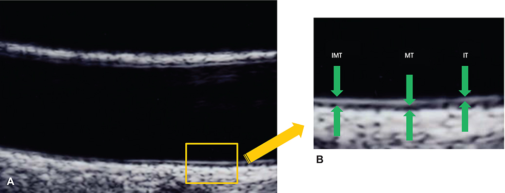
Fig. 1 The double line pattern and separate measurement of carotid intima-media thickness. Carotid IMT is defined as the distance between the luminal border of the intima and the outer border of media of carotid artery far wall on a B-mode ultrasound image. (A) True longitudinal plane simultaneously demonstrating double lines on the near and far walls of the common carotid artery. (B) Separately IMT, MT, and IT measured on far walls of the common carotid artery. IMT: intima-media thickness, MT: media thickness, IT: intima thickness. Modified with permission from Kim et al.21)
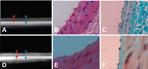
Fig. 2 Comparison of HEIT and IT. (A-C) Normotensive rats. (D-F) Spontaneously hypertensive rats SHR. (A) Measurement of HEIT and IMT on UBM images in normotensive rats. Red arrow indicates HEIT, and blue arrow indicates IMT. (B) The thin layer of endothelial cells is shown at the intimal area of the carotid artery. (C) Example of the thin layer of intima stained with MT staining. (D) Measurement of HEIT and IMT on UBM images in SHR. (E) The thick layer of endothelial cells is shown at the intimal area of the carotid artery. (F) Example of a thickened intimal layer with endothelial cells is shown with MT staining. HEIT: high-echogenic intimal thickening, IT: intimal thickness, IMT: intima-media thickness, MT: Masson's trichrome, UBM: ultrasound biomicroscope, SHR: spontaneously hypertensive rat.
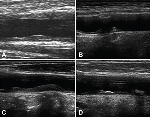
Fig. 3 Variable tissue characterization of carotid plaque. (A) Single or focal plaque on normal common carotid intima-media thickness. (B) Irregular surface plaque on carotid bulb. (C) Diffuse longitudinal and echolucent plaque on common carotid artery. (D) Calcified and heterogeneous plaque on common carotid artery.
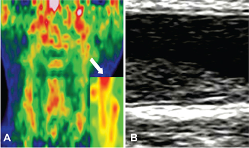
Fig. 4 Example of the image from integrated PET/CT device at transverse image and carotid ultrasonography. (A) Highly 18FDG uptake at left carotid artery. (B) Echolucent plaque was noted at left carotid artery. PET/CT: positron emission tomography-computed tomography.
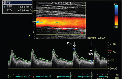
Fig. 5 Doppler findings of common carotid artery. PSV: peak systolic velocity, EDV: end-diastolic velocity.
Cited by 1 articles
-
Effect of Niacin on Carotid Atherosclerosis in Patients at Low-Density Lipoprotein-Cholesterol Goal but High Lipoprotein (a) Level: a 2-Year Follow-Up Study
Shinjeong Song, Chan Joo Lee, Jaewon Oh, Sungha Park, Seok-Min Kang, Sang-Hak Lee
J Lipid Atheroscler. 2019;8(1):58-66. doi: 10.12997/jla.2019.8.1.58.
Reference
-
1. Schmidt-Trucksass A, Grathwohl D, Schmid A, et al. Structural, functional, and hemodynamic changes of the common carotid artery with age in male subjects. Arterioscler Thromb Vasc Biol. 1999; 19:1091–1097.2. Schoning M, Walter J, Scheel P. Estimation of cerebral blood flow through color duplex sonography of the carotid and vertebral arteries in healthy adults. Stroke. 1994; 25:17–22.3. Pignoli P, Tremoli E, Poli A, Oreste P, Paoletti R. Intimal plus medial thickness of the arterial wall: a direct measurement with ultrasound imaging. Circulation. 1986; 74:1399–1406.4. Stein JH, Korcarz CE, Hurst RT, et al. Use of carotid ultrasound to identify subclinical vascular disease and evaluate cardiovascular disease risk: a consensus statement from the American Society of Echocardiography Carotid Intima-Media Thickness Task Force. Endorsed by the Society for Vascular Medicine. J Am Soc Echocardiogr. 2008; 21:93–111. quiz 189-90.5. Howard G, Sharrett AR, Heiss G, et al. Carotid artery intimal-medial thickness distribution in general populations as evaluated by B-mode ultrasound. ARIC Investigators. Stroke. 1993; 24:1297–1304.6. Polak JF, Pencina MJ, Pencina KM, O'Donnell CJ, Wolf PA, D'Agostino RB Sr. Carotid-wall intima-media thickness and cardiovascular events. N Engl J Med. 2011; 365:213–221.7. van der Meer IM, Bots ML, Hofman A, del Sol AI, van der Kuip DA, Witteman JC. Predictive value of noninvasive measures of atherosclerosis for incident myocardial infarction: the Rotterdam Study. Circulation. 2004; 109:1089–1094.8. Salonen JT, Salonen R. Ultrasonographically assessed carotid morphology and the risk of coronary heart disease. Arterioscler Thromb. 1991; 11:1245–1249.9. Naqvi TZ, Mendoza F, Rafii F, et al. High prevalence of ultrasound detected carotid atherosclerosis in subjects with low Framingham risk score: potential implications for screening for subclinical atherosclerosis. J Am Soc Echocardiogr. 2010; 23:809–815.10. Chuang SY, Bai CH, Cheng HM, et al. Common carotid artery end-diastolic velocity is independently associated with future cardiovascular events. Eur J Prev Cardiol. 2016; 23:116–124.11. Goff DC Jr, Lloyd-Jones DM, Bennett G, et al. 2013 ACC/AHA guideline on the assessment of cardiovascular risk: a report of the American College of Cardiology/American Heart Association Task Force on Practice Guidelines. JACC. 2014; 63:2935–2959.12. Naqvi TZ, Lee MS. Carotid intima-media thickness and plaque in cardiovascular risk assessment. JACC Cardiovasc Imaging. 2014; 7:1025–1038.13. Roman MJ, Naqvi TZ, Gardin JM, et al. Clinical application of noninvasive vascular ultrasound in cardiovascular risk stratification: a report from the American Society of Echocardiography and the Society of Vascular Medicine and Biology. J Am Soc Echocardiogr. 2006; 19:943–954.14. del Sol AI, Moons KG, Hollander M, et al. Is carotid intima-media thickness useful in cardiovascular disease risk assessment? The Rotterdam Study. Stroke. 2001; 32:1532–1538.15. Kanters SD, Algra A, van Leeuwen MS, Banga JD. Reproducibility of in vivo carotid intima-media thickness measurements: a review. Stroke. 1997; 28:665–671.16. Choi YS, Youn HJ, Youn JS, Park CS, Oh YS, Chung WS. Measurement of the intimal thickness of the carotid artery: comparison between 40 MHz ultrasound and histology in rats. Ultrasound Med Biol. 2009; 35:962–966.17. Bae JH, Kim WS, Rihal CS, Lerman A. Individual measurement and significance of carotid intima, media, and intima-media thickness by B-mode ultrasonographic image processing. Arterioscler Thromb Vasc Biol. 2006; 26:2380–2385.18. Gussenhoven EJ, Frietman PA, The SH, et al. Assessment of medial thinning in atherosclerosis by intravascular ultrasound. Am J Cardiol. 1991; 68:1625–1632.19. Nakashima Y, Chen YX, Kinukawa N, Sueishi K. Distributions of diffuse intimal thickening in human arteries: preferential expression in atherosclerosis-prone arteries from an early age. Virchows Arch. 2002; 441:279–288.20. Won HK, Kim WS, Kim KY, Hyun DW, Kwon TG, Bae JH. Increased carotid intima-media thickness in hypertensive patients is caused by increased medial thickness. Korean J Med. 2008; 75:179–185.21. Kim JH, Youn HJ, Kim GH, Moon KW, Yoo KD, Kim CM. The clinical significance of separate measurements of carotid arterial wall to assess the risk factor for atherosclerosis. J Cardiovasc Ultrasound. 2016; 24:48–54.22. Wikstrand J. Methodological considerations of ultrasound measurement of carotid artery intima-media thickness and lumen diameter. Clin Physiol Funct Imaging. 2007; 27:341–345.23. O'Leary DH, Polak JF, Kronmal RA, et al. Distribution and correlates of sonographically detected carotid artery disease in the Cardiovascular Health Study. The CHS Collaborative Research Group. Stroke. 1992; 23:1752–1760.24. Hlatky MA, Greenland P, Arnett DK, et al. Criteria for evaluation of novel markers of cardiovascular risk: a scientific statement from the American Heart Association. Circulation. 2009; 119:2408–2416.25. Den Ruijter HM, Peters SA, Anderson TJ, et al. Common carotid intima-media thickness measurements in cardiovascular risk prediction: a meta-analysis. JAMA. 2012; 308:796–803.26. Inaba Y, Chen JA, Bergmann SR. Carotid plaque, compared with carotid intima-media thickness, more accurately predicts coronary artery disease events: a meta-analysis. Atherosclerosis. 2012; 220:128–133.27. Peters SA, den Ruijter HM, Bots ML, Moons KG. Improvements in risk stratification for the occurrence of cardiovascular disease by imaging subclinical atherosclerosis: a systematic review. Heart. 2012; 98:177–184.28. Nambi V, Chambless L, He M, et al. Common carotid artery intima-media thickness is as good as carotid intima-media thickness of all carotid artery segments in improving prediction of coronary heart disease risk in the Atherosclerosis Risk in Communities (ARIC) study. Eur Heart J. 2012; 33:183–190.29. Kim G, Youn HJ, Choi YS, Jung HO, Chung WS, Kim CM. Is carotid artery evaluation necessary for primary prevention in asymptomatic high-risk patients without atherosclerotic cardiovascular disease? Clin Interv Aging. 2015; 10:1111–1119.30. Lee S, Cho GY, Kim HS, et al. Common carotid intima-media thickness as a risk factor for outcomes in Asian patients with acute ST-elevation myocardial infarction. Can J Cardiol. 2014; 30:1620–1626.31. Kim GH, Youn HJ, Choi YS, Jung HO, Chung WS, Kim CM. Carotid artery evaluation and coronary calcium score: which is better for the diagnosis and prevention of atherosclerotic cardiovascular disease? Int J Clin Exp Med. 2015; 8:18591–18600.32. Yu JS, Choi YS, Kim JY, et al. Carotid intima-media thickness is not related with clinical outcomes in young hypertensives. Clin Hypertens. 2015; 21:15.33. Azarpazhooh MR, Kazemi-Bajestani SM, Esmaeili H, et al. Cardiovascular risk factors and nutritional intake are not associated with ultrasound-defined increased carotid intima media thickness in individuals without a history of cardiovascular events. Int J Prev Med. 2014; 5:1412–1421.34. Thompson T, Shields KJ, Barinas-Mitchell E, Newman A, Sutton-Tyrrell K. Calcified carotid artery plaques predict cardiovascular outcomes in the elderly. J Hypertens. 2015; 33:810–817. discussion 817.35. Prabhakaran S, Singh R, Zhou X, Ramas R, Sacco RL, Rundek T. Presence of calcified carotid plaque predicts vascular events: the Northern Manhattan Study. Atherosclerosis. 2007; 195:e197–e201.36. Nambi V, Chambless L, Folsom AR, et al. Carotid intima-media thickness and presence or absence of plaque improves prediction of coronary heart disease risk: the ARIC (Atherosclerosis Risk In Communities) study. J Am Coll Cardiol. 2010; 55:1600–1607.37. Peters SA, Dogan S, Meijer R, et al. The use of plaque score measurements to assess changes in atherosclerotic plaque burden induced by lipid-lowering therapy over time: the METEOR study. J Atheroscler Thromb. 2011; 18:784–795.38. Plichart M, Celermajer DS, Zureik M, et al. Carotid intima-media thickness in plaque-free site, carotid plaques and coronary heart disease risk prediction in older adults. The Three-City Study. Atherosclerosis. 2011; 219:917–924.39. Kim JH, Youn HJ, Hong EJ, et al. Clinical significance of B-mode ultrasound of common carotid artery for prediction of severity of coronary artery disease: important parameters on hand measurement. Korean Circ J. 2005; 35:467–473.40. Choi YS, Youn HJ, Chung WB, et al. Uptake of F-18 FDG and ultrasound analysis of carotid plaque. J Nucl Cardiol. 2011; 18:267–272.41. Wahlgren CM, Zheng W, Shaalan W, Tang J, Bassiouny HS. Human carotid plaque calcification and vulnerability. Relationship between degree of plaque calcification, fibrous cap inflammatory gene expression and symptomatology. Cerebrovasc Dis. 2009; 27:193–200.42. Wasserman BA, Sharrett AR, Lai S, et al. Risk factor associations with the presence of a lipid core in carotid plaque of asymptomatic individuals using high-resolution MRI: the multi-ethnic study of atherosclerosis (MESA). Stroke. 2008; 39:329–335.43. Lal BK, Hobson RW 2nd, Pappas PJ, et al. Pixel distribution analysis of B-mode ultrasound scan images predicts histologic features of atherosclerotic carotid plaques. J Vasc Surg. 2002; 35:1210–1217.44. Sillesen H, Muntendam P, Adourian A, et al. Carotid plaque burden as a measure of subclinical atherosclerosis: comparison with other tests for subclinical arterial disease in the High Risk Plaque BioImage study. JACC Cardiovasc Imaging. 2012; 5:681–689.45. Khalil A, Huffman MD, Prabhakaran D, et al. Predictors of carotid intima-media thickness and carotid plaque in young Indian adults: the New Delhi birth cohort. Int J Cardiol. 2013; 167:1322–1328.46. Vasan RS. Biomarkers of cardiovascular disease: molecular basis and practical considerations. Circulation. 2006; 113:2335–2362.47. Cohen GI, Aboufakher R, Bess R, et al. Relationship between carotid disease on ultrasound and coronary disease on CT angiography. JACC Cardiovasc Imaging. 2013; 6:1160–1167.48. Ikeda N, Kogame N, Iijima R, Nakamura M, Sugi K. Impact of carotid artery ultrasound and ankle-brachial index on prediction of severity of SYNTAX score. Circ J. 2013; 77:712–716.49. Liang Y, Hou Y, Niu H, Lu M, Xue L, Sun Q. Correlation of high-sensitivity C-reactive protein and carotid plaques with coronary artery disease in elderly patients. Exp Ther Med. 2015; 10:275–278.50. Bai CH, Chen JR, Chiu HC, Pan WH. Lower blood flow velocity, higher resistance index, and larger diameter of extracranial carotid arteries are associated with ischemic stroke independently of carotid atherosclerosis and cardiovascular risk factors. J Clin Ultrasound. 2007; 35:322–330.51. Chuang SY, Bai CH, Chen JR, et al. Common carotid end-diastolic velocity and intima-media thickness jointly predict ischemic stroke in Taiwan. Stroke. 2011; 42:1338–1344.52. Chung H, Jung YH, Kim KH, et al. Carotid artery end-diastolic velocity and future cerebro-cardiovascular events in asymptomatic high risk patients. Korean Circ J. 2016; 46:72–78.
- Full Text Links
-
- Actions
-
Cited
- CITED
-
- Close
- Share
-
- Similar articles
-
- Speckle Tracking of Common Carotid Artery: A New Method for the Evaluation of Mechanical Vascular Function of Atherosclerosis
- Relationship Between Carotid and Coronary Atherosclerosis in the Elderly
- Neutrophil-to-Lymphocyte Ratio for Risk Assessment in Coronary Artery Disease and Carotid Artery Atherosclerosis
- Prevalence of Carotid Atherosclerosis in the Elderly
- Carotid ultrasound in patients with coronary artery disease
