J Rheum Dis.
2016 Dec;23(6):392-395. 10.4078/jrd.2016.23.6.392.
Case of Follicular Bronchiolitis in Rheumatoid Arthritis
- Affiliations
-
- 1Department of Internal Medicine, Presbyterian Medical Center, Jeonju, Korea.
- 2Department of Pathology, Presbyterian Medical Center, Jeonju, Korea.
- 3Division of Allergy and Pulmonology, Department of Internal Medicine, Presbyterian Medical Center, Jeonju, Korea. usdina@naver.com
- KMID: 2364694
- DOI: http://doi.org/10.4078/jrd.2016.23.6.392
Abstract
- Follicular bronchiolitis is an uncommon bronchiolar disorder that is characterized by the presence of hyperplastic lymphoid follicles with reactive germinal centers. The condition is associated with connective tissue diseases such as rheumatoid arthritis, Sjögren's syndrome, and immunodeficiency disorders. A 56-year-old man with rheumatoid arthritis was admitted to hospital with a progressively enlarging pulmonary nodule in the left upper lobe. A follow-up contrast tomography scan showed that the nodule had increased in size from 4.2 mm to 6.3 mm over a 3 month period. An open lung biopsy was performed to establish a definite pathologic diagnosis of the pulmonary nodule, which was suspected to be a lung malignancy. The nodule was diagnosed as follicular bronchiolitis based on the histopathology findings. We describe a patient with follicular bronchiolitis that was confirmed by an open lung biopsy, and is believed to have had rheumatoid involvement.
MeSH Terms
Figure
-
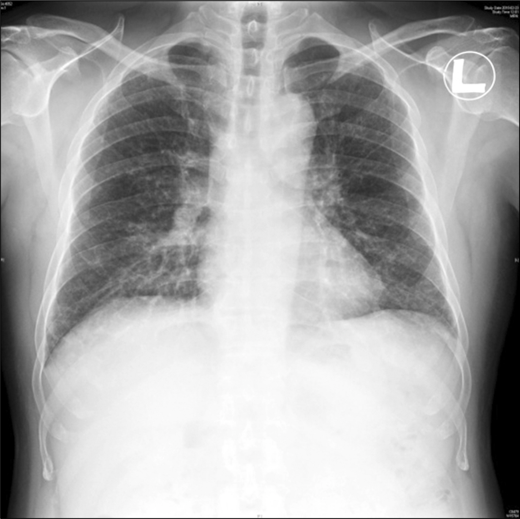
Figure 1. The chest radiograph revealed linear densities and small nodules bilaterally in the lower lung fields.
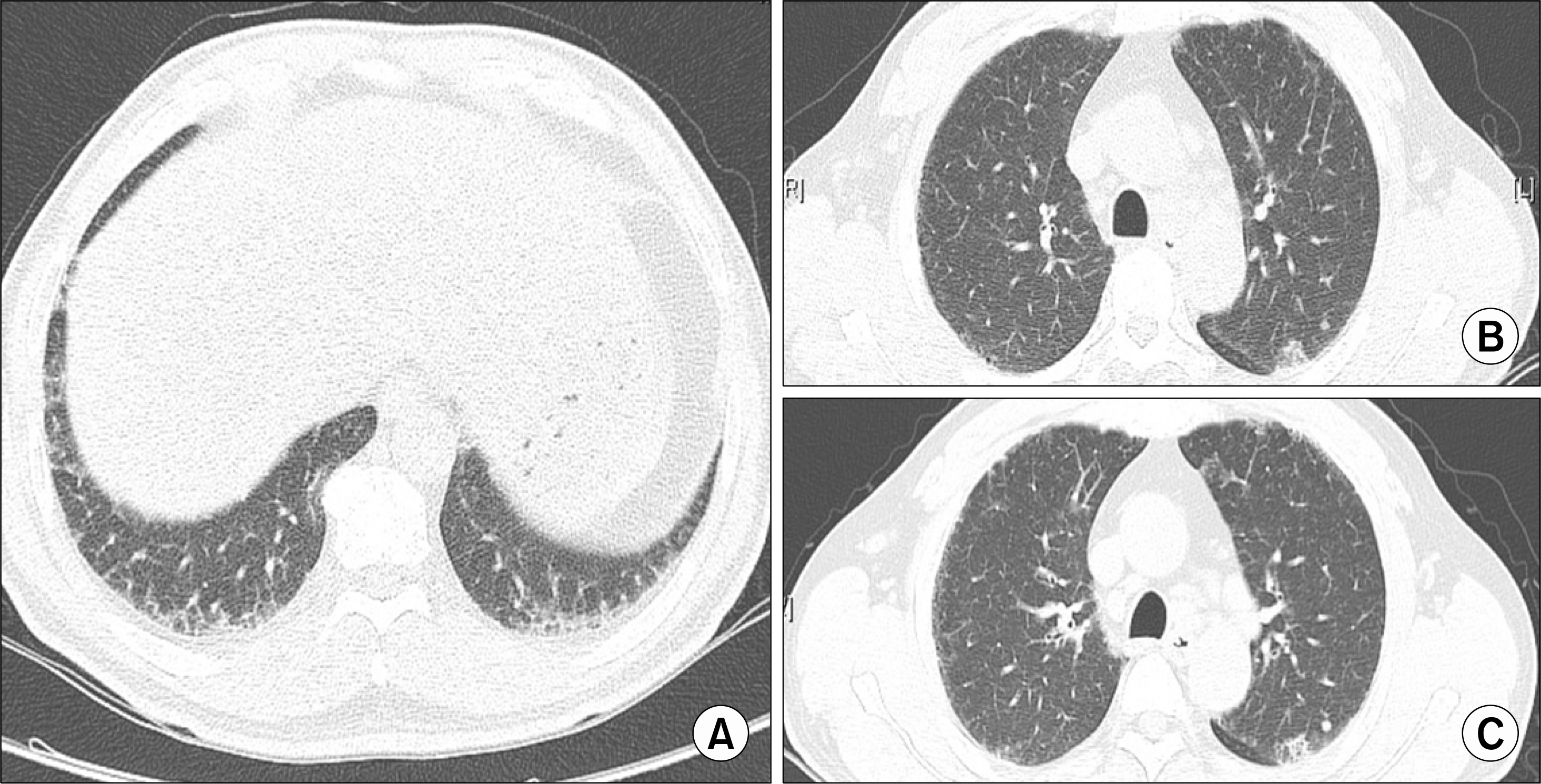
Figure 2. Chest computed tomography revealed (A) linear, reticular densities and ground glass opacities in the bilateral lower lung fields, as well as (B and C) a slightly increased nodule (that had increased in size from 4.2 mm to 6.3 mm) in the apicoposterior segment of the left upper lobe.
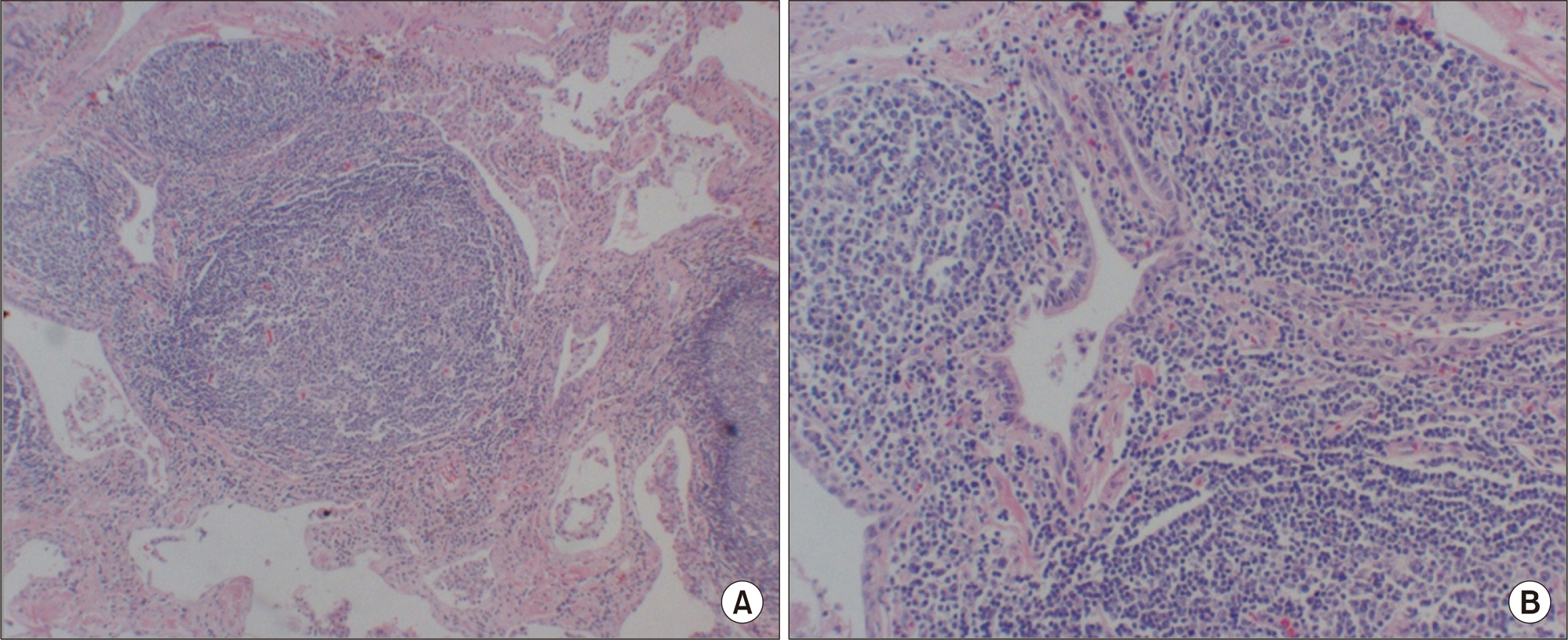
Figure 3. Histologic specimen showing (A) hyperplasia of the lymphoid follicle (H&E, ×40) and (B) a benign hyperplastic germinal center (H&E, ×100).
Reference
-
1. Archontogeorgis K, Steiropoulos P, Tzouvelekis A, Nena E, Bouros D. Lung cancer and interstitial lung diseases: a systematic review. Pulm Med. 2012; 2012; 315918.
Article2. Nicholson AG, Wotherspoon AC, Diss TC, Hansell DM, Du Bois R, Sheppard MN, et al. Reactive pulmonary lymphoid disorders. Histopathology. 1995; 26:405–12.
Article3. Yousem SA, Colby TV, Carrington CB. Follicular bronchitis/bronchiolitis. Hum Pathol. 1985; 16:700–6.
Article4. Aerni MR, Vassallo R, Myers JL, Lindell RM, Ryu JH. Follicular bronchiolitis in surgical lung biopsies: clinical implications in 12 patients. Respir Med. 2008; 102:307–12.
Article5. Lee YJ, Park JH, Cho GJ, Lee BC, Kim DS, Suh YL, et al. A case of follicular bronchitis/bronchiolitis. Korean J Med. 1993; 45:795–801.6. Kim MS, Lim SC, Kim YH, Na KJ, Kim KS, Kwon KY, et al. a case report of localized form of follicular bronchitis/bronchiolitis with fibrosis. Tuberc Respir Dis. 1998; 45:191–6.
Article7. Howling SJ, Hansell DM, Wells AU, Nicholson AG, Flint JD, Müller NL. Follicular bronchiolitis: thin-section CT and histologic findings. Radiology. 1999; 212:637–42.
Article8. Kim EJ, Collard HR, King TE Jr. Rheumatoid arthritis-associated interstitial lung disease: the relevance of histopathologic and radiographic pattern. Chest. 2009; 136:1397–405.9. American Thoracic Society; European Respiratory Society. American Thoracic Society/European Respiratory Society International Multidisciplinary Consensus Classification of the Idiopathic Interstitial Pneumonias. This joint statement of the American Thoracic Society (ATS), and the European Respiratory Society (ERS) was adopted by the ATS board of directors, June 2001 and by the ERS Executive Committee, June 2001. Am J Respir Crit Care Med. 2002; 165:277–304.
- Full Text Links
-
- Actions
-
Cited
- CITED
-
- Close
- Share
-
- Similar articles
-
- A Case Report of Localized Form of Follicular Bronchitis/Bronchiolitis with Fibrosis
- A Case of Bronchiolitis Obliterans Organizing Pneumonia anteceded by Rheumatoid Arthritis
- Follicular Bronchiolitis Mimicking Lung Cancer in a Patient with Primary Sjögren's Syndrome
- Cytokines in rheumatoid arthritis
- Clinical significance of rheumatoid factor in juvenile rheumatoid arthritis
