Enhancement of Gastric Ulcer Healing and Angiogenesis by Hepatocyte Growth Factor Gene Mediated by Attenuated Salmonella in Rats
- Affiliations
-
- 1Department of Clinical Laboratory Medicine, Lanzhou General Hospital of Lanzhou Military Region, People's Liberation Army, Key Laboratory of Stem Cell and Gene Drug in Gansu Province, Lanzhou, China. haxiaoqin2013@163.com
- KMID: 2364161
- DOI: http://doi.org/10.3346/jkms.2017.32.2.186
Abstract
- The present study developed an oral hepatocyte growth factor (HGF) gene therapy strategy for gastric ulcers treatment. An attenuated Salmonella typhimurium that stably expressed high HGF (named as TPH) was constructed, and the antiulcerogenic effect of TPH was evaluated in a rat model of gastric ulcers that created by acetic acid subserosal injection. From day 5 after injection, TPH (1 × 10â¹ cfu), vehicle (TP, 1 × 10â¹ cfu), or sodium bicarbonate (model control) was administered orally every alternate day for three times. Then ulcer size was measured at day 21 after ulcer induction. The ulcer area in TPH-treated group was 10.56 ± 3.30 mm², which was smaller when compared with those in the TP-treated and model control groups (43.47 ± 4.18 and 56.25 ± 6.38 mm², respectively). A higher level of reepithelialization was found in TPH-treated group and the crawling length of gastric epithelial cells was significantly longer than in the other two groups (P < 0.05). The microvessel density in the ulcer granulation tissues of the TPH-treated rats was 39.9 vessels/mm², which was greater than in the TP-treated and model control rats, with a significant statistical difference. These results suggest that TPH treatment significantly accelerates the healing of gastric ulcers via stimulating proliferation of gastric epithelial cells and enhancing angiogenesis on gastric ulcer site.
MeSH Terms
Figure
-
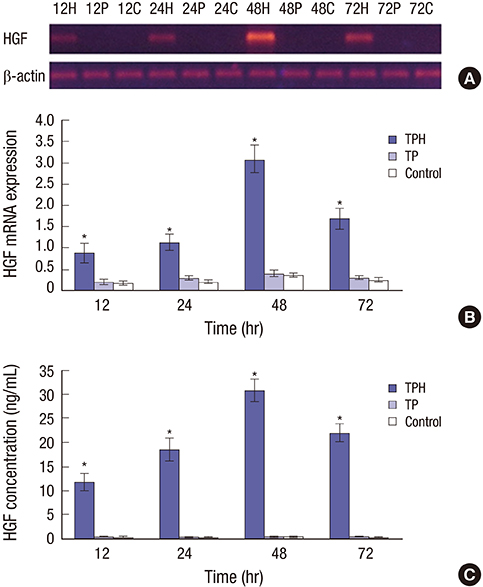
Fig. 1 HGF expression in the GES-1 cells after transfection with TPH or TP at different time points. (A) RT-PCR products of HGF and β-actin in the agar gel electrophoresis. (B) The statistical results of RT-PCR of HGF in the GES-1 cells. TPH group: 3 × 10⁷cfu TPH strain was added into the wells of a 6-well plate; TP group: 3 × 10⁷cfu TP strain was added into the wells of a 6-well plate; control group: PBS (10 μL/well) was added into the wells of a 6-well plate; 12, 24, 48, and 72 hours indicate transfected TPH or TP or PBS for 12, 24, 48, and 72 hours, respectively. (C) Results from an ELISA of HGF concentration in cell supernatants. Cell supernatants were collected at different times after transfection, and the HGF levels were detected by ELISA. TPH group: 3 × 10⁷ cfu TPH strain was added into the wells of a 6-well plate; TP group: 3 × 10⁷ cfu TP strain was added into the wells of a 6-well plate; control group: PBS (10 μL/well) was added into the wells of a 6-well plate; 12, 24, 48, and 72 hours indicate the HGF levels from supernatant of cells transfected by TPH or TP or PBS for 12, 24, 48, and 72 hours, respectively. GES-1 = gastric epithelial cells, TPH = attenuated Salmonella typhimurium strain carrying a eukaryotic expression vector encodes the human HGF gene, TP = attenuated Salmonella typhimurium strain with a eukaryotic expression vector, HGF = hepatocyte growth factor, RT-PCR = reverse transcription-polymerase chain reaction, PBS = phosphate buffer solution, ELISA = enzyme-linked immunosorbent assay. *P < 0.01, TPH group vs. TP and control groups.
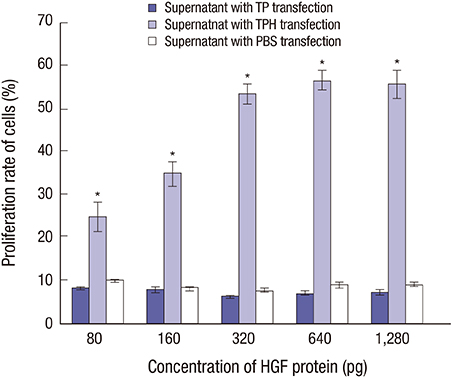
Fig. 2 TPH induced GES-1 cell proliferation. TPH transfected the GES-1 cells. After incubation at 37°C for 48 hours, cell proliferation was measured by an MTT assay. TPH group: 3 × 10³ cells/well (transfected by TPH strain) in a 96-well plate incubated at 37°C for 48 hours; TP group: 3 × 10³ cells/well (transfected by TP strain) in a 96-well plate incubated at 37°C for 48 hours; control group: 3 × 10³ cells/well (transfected by PBS) in a 96-well plate incubated at 37°C for 48 hours. GES-1 = gastric epithelial cells, MTT = 2-(4,5-dimethyltriazol-2-yl)-2,5-diphenyl tetrazolium bromide, TPH = attenuated Salmonella typhimurium strain carrying a eukaryotic expression vector encodes the human HGF gene, TP = attenuated Salmonella typhimurium strain with a eukaryotic expression vector, PBS = phosphate buffer solution. *P < 0.01, TPH group vs. TP and control groups.
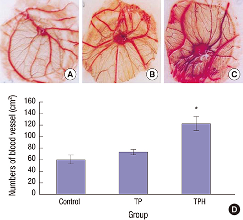
Fig. 3 Angiogenesis activity of the HGF expression product on the chick CAM. (A) Control group: only a methylcellulose disk on CAM. (B) TP group: a disk with same volume supernatant from TP-transfected supernatant on CAM. (C) TPH group: a disk with the HGF expression product (300 pg) on CAM, from TPH-transfected supernatant. (D) The number of blood vessels was counted under a light microscope, and was found to be significantly fewer in expression supernatant from the TP-transfected and control groups than in the HGF expression product from the TPH-transfected group (P < 0.05). HGF = hepatocyte growth factor, CAM = chorioallantoic membrane, TP = attenuated Salmonella typhimurium strain with a eukaryotic expression vector, TPH = attenuated Salmonella typhimurium strain carrying a eukaryotic expression vector encodes the human HGF gene. *P < 0.05, TPH group vs. TP and control groups.
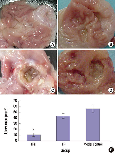
Fig. 4 Gross observation of gastric mucosa on day 21 after induced ulcer treatment. (A) Normal control group: the gastric mucosae from normal rats were smooth and intact with no ulceration. (B) TPH treatment group: treated with (1 × 10⁹) cfu TPH for acetic acid–induced gastric ulcer rats by gavage, the gastric mucosae of the TPH-treatment group were similar to the normal rats. The gastric mucosal lesions were smaller and shallow, and the regenerative granulation tissues were observed in ulcers. (C) Model control group: treated with 0.5-mL1.19M NaHCO3 for acetic acid–induced gastric ulcer rats by gavage. (D) TP treatment group: treated with 1 × 10⁹ cfu TP for acetic acid–induced gastric ulcer rats by gavage. In the model control and TP groups, enlarged, deepened ulcers with severe adhesions to adjacent tissues were observed, the mucosa underwent necrosis, ulcerations, and had a dark yellowish membrane-like coating. The adjacent mucosa had obvious hyperemia and edema. (E) The statistical results of the mean ulcer area size (mm2). The size of the ulcer area in 21 days after TPH treatment was smaller than that in the other two groups. TPH = attenuated Salmonella typhimurium strain carrying a eukaryotic expression vector encodes the human HGF gene, TP = attenuated Salmonella typhimurium strain with a eukaryotic expression vector. *P < 0.01, TPH group vs. TP and control groups.
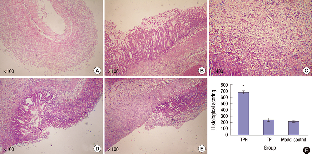
Fig. 5 Histological observations of the gastric mucosa. (A) Normal control group: Clear glands and intact mucosa with abundant submucosal vessels were observed (H&E stain × 100). (B) TPH treatment group: Increased number of glands and submucosal granulation tissue regeneration were observed in the bases of ulcers along with regenerated capillaries. The gastric mucosa significantly healed (H&E stain × 100). (C) TPH treatment group: Increased number of glands and submucosal granulation tissue regeneration were observed in the bases of ulcers along with regenerated capillaries. The gastric mucosa significantly healed (H&E stain × 400). (D) TP-treated group: Severe mucosal anabrosis and depletion with ulceration were observed. The reepithelialization of gastric mucosa increased in the TP-treated group than in the model control group; however, no significant histological difference was found between the TP and the model control groups (H&E stain × 100). (E) Model control group: Severe mucosal anabrosis and depletion with ulceration were observed (H&E stain × 100). (F) The histological scoring for the crawling length of gastric epithelial cells from three groups. After treatment with TPH, the crawling length of gastric epithelial cells was significantly longer than in the TP and model control groups (P < 0.05). No statistical significance was observed in the mucosal epithelial crawling length between the TP and the model control groups after treatments (P > 0.05). H&E stain = hematoxylin-eosin stain, TPH = attenuated Salmonella typhimurium strain carrying a eukaryotic expression vector encodes the human HGF gene, TP = attenuated Salmonella typhimurium strain with a eukaryotic expression vector. *P < 0.01, TPH group vs. TP and control groups.
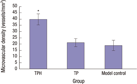
Fig. 6 Quantification of MVD on day 21 in granulation tissue from the ulcer beds by immunoreaction with CD34. Data are presented as mean ± standard error (n = 50). The MVD in the TPH-treated group increased compared to the TP (a recombinant attenuated Salmonella strain carrying a eukaryotic expression vector)-treated and model control groups, with a significant statistical difference. The MVD in the TP-treated group was not significantly different compared with the model control group. MVD = microvascular density, TPH = attenuated Salmonella typhimurium strain carrying a eukaryotic expression vector encodes the human HGF gene, TP = attenuated Salmonella typhimurium strain with a eukaryotic expression vector. *P < 0.01, TPH group vs. TP and control groups.
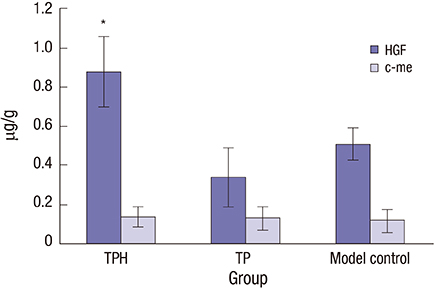
Fig. 7 HGF and c-Met protein expression in gastric mucosa by ELISA. On day 21, the expression of HGF protein in gastric ulcer was significantly higher in the TPH-treated group than in the TP-treated and model control groups. However, the protein expression of c-Met after 21 days of TPH treatment was not significantly different from the TP-treated and model control groups. HGF = hepatocyte growth factor, ELISA = enzyme-linked immunosorbent assay, TPH = attenuated Salmonella typhimurium strain carrying a eukaryotic expression vector encodes the human HGF gene, TP = attenuated Salmonella typhimurium strain with a eukaryotic expression vector. *P < 0.01, TPH group vs. TP and control groups.
Reference
-
1. Tanaka T, Arai M, Minemura S, Oyamada A, Saito K, Jiang X, Tsuboi M, Sazuka S, Maruoka D, Matsumura T, et al. Expression level of sonic hedgehog correlated with the speed of gastric mucosa regeneration in artificial gastric ulcers. J Gastroenterol Hepatol. 2014; 29:736–741.2. Kangwan N, Park JM, Kim EH, Hahm KB. Quality of healing of gastric ulcers: natural products beyond acid suppression. World J Gastrointest Pathophysiol. 2014; 5:40–47.3. Harada S, Takeuchi T, Edogawa S, Ota K, Kojima Y, Higuchi K. The availability of prostaglandin derivatives in a treatment and prevention for gastrointestinal mucosal injury. Nihon Rinsho. 2015; 73:1153–1158.4. Sugiyama T. Mucosal protective drugs. Nihon Rinsho. 2015; 73:1147–1152.5. Samsonov AA, Grechushnikov VB, Andreev DN, Iurenev GL, Korovina TI, Lezhneva IA, Maev IV. Pharmacoeconomic evaluation of treatment in patients with Helicobacter pylori-associated diseases. Ter Arkh. 2014; 86:56–61.6. Szabo S, Vincze A. Growth factors in ulcer healing: lessons from recent studies. J Physiol Paris. 2000; 94:77–81.7. Takahashi M, Takada H, Takagi K, Kataoka S, Soma R, Kuwayama H. Gastric restitution is inhibited by dexamethasone, which is reversed by hepatocyte growth factor and rebamipide. Aliment Pharmacol Ther. 2003; 18:Suppl 1. 126–132.8. Sato T, Amano H, Ito Y, Eshima K, Minamino T, Ae T, Katada C, Ohno T, Hosono K, Suzuki T, et al. Vascular endothelial growth factor receptor 1 signaling facilitates gastric ulcer healing and angiogenesis through the upregulation of epidermal growth factor expression on VEGFR1+CXCR4+ cells recruited from bone marrow. J Gastroenterol. 2014; 49:455–469.9. Uematsu Y, Fujise N, Kohsaka K, Masunaga H, Higashio K. Effective administration route for the deleted form of hepatocyte growth factor to exert its pharmacological effects. J Pharm Sci. 1999; 88:131–135.10. Qiu L, Wang X, Hao H, Mu G, Dang R, Wang J, Zhang S, Du E, Yang Z. Oral administration of attenuated Salmonella typhimurium containing a DNA vaccine against rabbit haemorrhagic disease. J Virol Methods. 2013; 188:108–113.11. Sambrook J, Fritsch EF, Maniatis T. Molecular Cloning: a Laboratory Manual. New York , NY: Cold Spring Harbor Laboratory Press;1989.12. Bai LY, Liang AX, Zhang J, Yang FF, Han L, Huo LJ, Yang LG. Effects of immunization against a DNA vaccine encoding somatostatin gene (pGM-CSF/SS) by attenuated Salmonella typhimurium on growth, reproduction and lactation in female mice. Theriogenology. 2011; 75:155–163.13. Dusseau JW, Hutchins PM. Microvascular responses to chronic hypoxia by the chick chorioallantoic membrane: a morphometric analysis. Microvasc Res. 1989; 37:138–147.14. Okabe S, Amagase K. An overview of acetic acid ulcer models--the history and state of the art of peptic ulcer research. Biol Pharm Bull. 2005; 28:1321–1341.15. Kang JM, Kim N, Kim B, Kim JH, Lee BY, Park JH, Lee MK, Lee HS, Kim JS, Jung HC, et al. Enhancement of gastric ulcer healing and angiogenesis by cochinchina Momordica seed extract in rats. J Korean Med Sci. 2010; 25:875–881.16. Weidner N, Gasparini G. Determination of epidermal growth factor receptor provides additional prognostic information to measuring tumor angiogenesis in breast carcinoma patients. Breast Cancer Res Treat. 1994; 29:97–107.17. Li J, Zheng CQ, Li Y, Yang C, Lin H, Duan HG. Hepatocyte growth factor gene-modified mesenchymal stem cells augment sinonasal wound healing. Stem Cells Dev. 2015; 24:1817–1830.18. Nakamura T, Nawa K, Ichihara A. Partial purification and characterization of hepatocyte growth factor from serum of hepatectomized rats. Biochem Biophys Res Commun. 1984; 122:1450–1459.19. Kaido T, Imamura M. Hepatocyte growth factor: clinical implications in hepatobiliary pancreatic surgery. J Hepatobiliary Pancreat Surg. 2001; 8:65–75.20. Li Z, Yin PH, Yang SS, Li QY, Chang T, Fang L, Shi LX, Fang GE. Recombinant attenuated Salmonella typhimurium carrying a plasmid co-expressing ENDO-VEGI151 and survivin siRNA inhibits the growth of breast cancer in vivo. Mol Med Rep. 2013; 7:1215–1222.21. van Golde J, Mulder T, Blanco CE. Changes in mean chorioallantoic artery blood flow and heart rate produced by hypoxia in the developing chick embryo. Pediatr Res. 1997; 42:293–298.22. Staton CA, Stribbling SM, Tazzyman S, Hughes R, Brown NJ, Lewis CE. Current methods for assaying angiogenesis in vitro and in vivo. Int J Exp Pathol. 2004; 85:233–248.23. Nowak-Sliwinska P, Ballini JP, Wagnières G, van den Bergh H. Processing of fluorescence angiograms for the quantification of vascular effects induced by anti-angiogenic agents in the CAM model. Microvasc Res. 2010; 79:21–28.24. Favia G, Mariggio MA, Maiorano F, Cassano A, Capodiferro S, Ribatti D. Accelerated wound healing of oral soft tissues and angiogenic effect induced by a pool of aminoacids combined to sodium hyaluronate (AMINOGAM). J Biol Regul Homeost Agents. 2008; 22:109–116.25. Takagi K, Okabe S, Saziki R. A new method for the production of chronic gastric ulcer in rats and the effect of several drugs on its healing. Jpn J Pharmacol. 1969; 19:418–426.26. de-Faria FM, Almeida AC, Luiz-Ferreira A, Dunder RJ, Takayama C, da Silva MS, da Silva MA, Vilegas W, Rozza AL, Pellizzon CH, et al. Mechanisms of action underlying the gastric antiulcer activity of the Rhizophora mangle L. J Ethnopharmacol. 2012; 139:234–243.
- Full Text Links
-
- Actions
-
Cited
- CITED
-
- Close
- Share
-
- Similar articles
-
- Enhancement of Gastric Ulcer Healing and Angiogenesis by Cochinchina Momordica Seed Extract in Rats
- Therapeutic Angiogenesis with Somatic Stem Cell Transplantation
- The Prognostic Value of Tumor Angiogenesis, Hepatocyte Growth Factor and c-met Expression in Renal Cell Carcinoma
- Changes of the Serum Hepatocyte Growth Factor Levels after Hepatectomy
- Expression of Epidermal Growth Factor at Ulcer Margin and Antral Mucosa in Patients with Gastric Ulcer
