J Korean Med Sci.
2016 Feb;31(Suppl 1):S69-S74. 10.3346/jkms.2016.31.S1.S69.
Radiation Dose from Whole-Body F-18 Fluorodeoxyglucose Positron Emission Tomography/Computed Tomography: Nationwide Survey in Korea
- Affiliations
-
- 1Department of Nuclear Medicine, Seoul National University Hospital, Seoul, Korea. paengjc@snu.ac.kr
- 2Department of Molecular Medicine and Biopharmaceutical Science, Graduate school of Convergence Science and Technology, Seoul National University, Seoul, Korea.
- 3Institute of Radiation Medicine, Medical Research Center, Seoul National University College of Medicine, Seoul, Korea.
- KMID: 2363383
- DOI: http://doi.org/10.3346/jkms.2016.31.S1.S69
Abstract
- The purpose of this study was to estimate average radiation exposure from 18F-fluorodeoxyglucose (FDG) positron emission tomography/computed tomography (PET/CT) examinations and to analyze possible factors affecting the radiation dose. A nation-wide questionnaire survey was conducted involving all institutions that operate PET/CT scanners in Korea. From the response, radiation doses from injected FDG and CT examination were calculated. A total of 105 PET/CT scanners in 73 institutions were included in the analysis (response rate of 62.4%). The average FDG injected activity was 310 +/- 77 MBq and 5.11 +/- 1.19 MBq/kg. The average effective dose from FDG was estimated to be 5.89 +/- 1.46 mSv. The average CT dose index and dose-length product were 4.60 +/- 2.47 mGy and 429.2 +/- 227.6 mGycm, which corresponded to 6.26 +/- 3.06 mSv. The radiation doses from FDG and CT were significantly lower in case of newer scanners than older ones (P < 0.001). Advanced PET technologies such as time-of-flight acquisition and point-spread function recovery were also related to low radiation dose (P < 0.001). In conclusion, the average radiation dose from FDG PET/CT is estimated to be 12.2 mSv. The radiation dose from FDG PET/CT is reduced with more recent scanners equipped with image-enhancing algorithms.
MeSH Terms
Figure
-
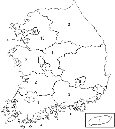
Fig. 1 Geometric distribution of the 73 institutions included in this survey.
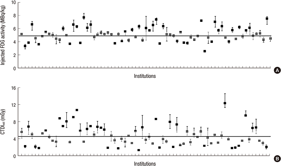
Fig. 2 Distribution of injected FDG activity and CTDIvol values of CT examinations in each institution. (A) Median value of injected FDG activity is 5.00 MBq/kg (25th–75th percentile, 4.24–5.62 MBq/kg; gray box). (B) Median value of CTDIvol is 4.10 mGy (25th–75th percentile, 2.80–5.96 mGy; gray box).
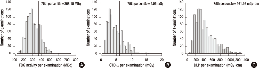
Fig. 3 Distribution of injected FDG activity (A), CTDIvol (B), and DLP (C) of each PET/CT examination.
Reference
-
1. Bly R, Jahnen A, Järvinen H, Olerud H, Vassileva J, Vogiatzi S. Collective effective dose in Europe from X-ray and nuclear medicine procedures. Radiat Prot Dosimetry. 2015; 165:129–132.2. Lee MC, Oh SW, Chung JK, Lee DS. The current status and future perspectives of nuclear medicine in Korea. Nucl Med Mol Imaging. 2010; 44:95–101.3. Korean Society of Nuclear Medicine. Annual statistics of nuclear medicine procedures in Korea. accessed on 1 September 2015. Available at http://www.ksnm.or.kr/education/sub2_5.php.4. ICRP. Radiation dose to patients from radiopharmaceuticals. Addendum 3 to ICRP Publication 53. ICRP Publication 106. Approved by the Commission in October 2007. Ann ICRP. 2008; 38:1–197.5. Stamm G, Nagel HD. CT-expo--a novel program for dose evaluation in CT. Rofo. 2002; 174:1570–1576.6. ICRP. Recommendations of the International Commision on Radiological Protection. ICRP Publication 60. Ann ICRP. 1991; 21:1–201.7. Linet MS, Slovis TL, Miller DL, Kleinerman R, Lee C, Rajaraman P, Berrington de Gonzalez A. Cancer risks associated with external radiation from diagnostic imaging procedures. CA Cancer J Clin. 2012; 62:75–100.8. Jahnen A, Järvinen H, Olerud H, Vassilieva J, Vogiatzi S, Shannoun F, Bly R. Analysis of factors correlating with medical radiological examination frequencies. Radiat Prot Dosimetry. 2015; 165:133–136.9. Health Insurance Review and Assessment Service of Korea. Statistics of medical procedures. accessed on 1 September 2015. Available at https://www.hira.or.kr/rd/dissdic/infoMdfeeList.do?pgmid=HIRAA020044030000.10. Etard C, Celier D, Roch P, Aubert B. National survey of patient doses from whole-body FDG PET-CT examinations in France in 2011. Radiat Prot Dosimetry. 2012; 152:334–338.11. Avramova-Cholakova S, Ivanova S, Petrova E, Garcheva M, Vassileva J. Patient doses from PET-CT procedures. Radiat Prot Dosimetry. 2015; 165:430–433.12. Rausch I, Bergmann H, Geist B, Schaffarich M, Hirtl A, Hacker M, Beyer T. Variation of system performance, quality control standards and adherence to international FDG-PET/CT imaging guidelines. A national survey of PET/CT operations in Austria. Nucl Med (Stuttg). 2014; 53:242–248.13. Boellaard R, O’Doherty MJ, Weber WA, Mottaghy FM, Lonsdale MN, Stroobants SG, Oyen WJ, Kotzerke J, Hoekstra OS, Pruim J, et al. FDG PET and PET/CT: EANM procedure guidelines for tumour PET imaging: version 1.0. Eur J Nucl Med Mol Imaging. 2010; 37:181–200.14. Boellaard R, Delgado-Bolton R, Oyen WJ, Giammarile F, Tatsch K, Eschner W, Verzijlbergen FJ, Barrington SF, Pike LC, Weber WA, et al. FDG PET/CT: EANM procedure guidelines for tumour imaging: version 2.0. Eur J Nucl Med Mol Imaging. 2015; 42:328–354.15. Karp JS, Surti S, Daube-Witherspoon ME, Muehllehner G. Benefit of time-of-flight in PET: experimental and clinical results. J Nucl Med. 2008; 49:462–470.16. Akamatsu G, Ishikawa K, Mitsumoto K, Taniguchi T, Ohya N, Baba S, Abe K, Sasaki M. Improvement in PET/CT image quality with a combination of point-spread function and time-of-flight in relation to reconstruction parameters. J Nucl Med. 2012; 53:1716–1722.17. Boellaard R, Oyen WJ, Hoekstra CJ, Hoekstra OS, Visser EP, Willemsen AT, Arends B, Verzijlbergen FJ, Zijlstra J, Paans AM, et al. The Netherlands protocol for standardisation and quantification of FDG whole body PET studies in multi-centre trials. Eur J Nucl Med Mol Imaging. 2008; 35:2320–2333.18. Graham MM, Wahl RL, Hoffman JM, Yap JT, Sunderland JJ, Boellaard R, Perlman ES, Kinahan PE, Christian PE, Hoekstra OS, et al. Summary of the UPICT protocol for 18F-FDG PET/CT imaging in oncology clinical trials. J Nucl Med. 2015; 56:955–961.19. ICRP. International Commission on Radiological Protection, Committee I. The evaluation of risks from radiation. Health Phys. 1966; 12:239–302.20. European Commission. Group of Advisers on the Ethical Implications of Biotechnology. Report to the European Commission: ethical aspects of cloning techniques. Politics Life Sci. 1997; 16:309–312.21. Botros GM, Smart RC, Towson JE. Diagnostic reference activities for nuclear medicine procedures in Australia and New Zealand derived from the 2008 survey. ANZ Nucl Med. 2009; 40:2–11.22. Korpela H, Bly R, Vassileva J, Ingilizova K, Stoyanova T, Kostadinova I, Slavchev A. Recently revised diagnostic reference levels in nuclear medicine in Bulgaria and in Finland. Radiat Prot Dosimetry. 2010; 139:317–320.
- Full Text Links
-
- Actions
-
Cited
- CITED
-
- Close
- Share
-
- Similar articles
-
- Whole Body Positron Emission Tomography/Computed Tomography
- F-18 fluorodeoxyglucose positron emission tomography/computed tomography in the infection of heart
- Fluorine-18 Fluorodeoxyglucose Positron Emission Tomography/Computed Tomography Findings of Post Traumatic Lymphangioma in a Young Adult Male
- The Use of PET in Esophageal Cancer
- Transient ¹â¸F-Fluorodeoxyglucose Activity on PET/CT of Herniation Pit in Thyroid Cancer Patient: A Case Report
