Ann Lab Med.
2015 May;35(3):321-328. 10.3343/alm.2015.35.3.321.
Interlaboratory Comparison of the Results of Lifecodes LSA Class I and Class II Single Antigen Kits for Human Leukocyte Antigen Antibody Detection
- Affiliations
-
- 1Department of Laboratory Medicine, Seoul St. Mary's Hospital, College of Medicine, The Catholic University of Korea, Seoul, Korea.
- 2Department of Laboratory Medicine, Seoul National University College of Medicine, Seoul, Korea. eysong1@snu.ac.kr
- 3Department of Laboratory Medicine, Seoul National University Bundang Hospital, Seongnam, Korea.
- 4Department of Laboratory Medicine and Genetics, Samsung Medical Center, Sungkyunkwan University School of Medicine, Seoul, Korea.
- 5Department of Laboratory Medicine, Severance Hospital, Yonsei University College of Medicine, Seoul, Korea.
- 6Department of Molecular Medicine and Biopharmaceutical Sciences, Graduate School of Convergence Science and Technology and College of Medicine, Medical Research Center, Seoul National University, Seoul, Korea.
- KMID: 2363204
- DOI: http://doi.org/10.3343/alm.2015.35.3.321
Abstract
- BACKGROUND
Although single antigen bead assays (SAB) are approved qualitative tests, the median fluorescence intensity (MFI) values obtained from SAB are frequently used in combination with quantitative significances for diagnostic purposes. To gauge the reproducibility of SAB results, we assessed the interlaboratory variability of MFI values using identical kits with reagents from the same lot and the manufacturer's protocol.
METHODS
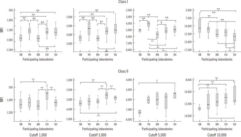
Six serum samples containing HLA-specific antibodies were analyzed at five laboratories by using Lifecodes LSA Class I and Class II SAB kits (Immucor, USA) from the same lot, according to the manufacturer's protocol. We analyzed the concordance of qualitative results according to distinct MFI cutoffs (1,000, 3,000, 5,000, and 10,000), and the correlation of quantitative MFI values obtained by the participating laboratories. The CV for MFI values were analyzed and grouped by mean MFI values from the five laboratories (<1,000; 1,000-2,999; 3,000-4,999; 5,000-9,999; and > or =10,000).
RESULTS
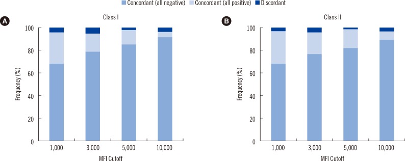
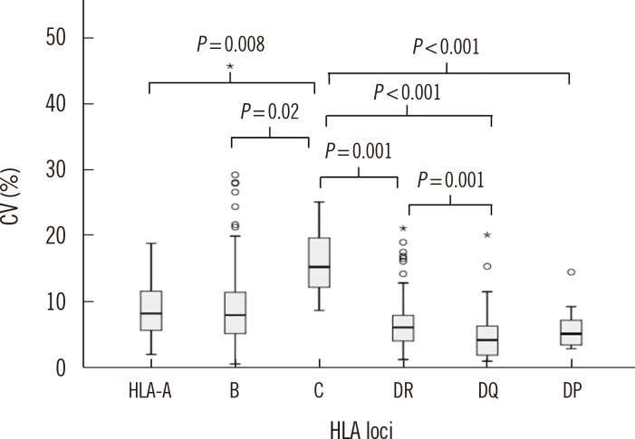
The categorical results obtained from the five laboratories exhibited concordance rates of 96.0% and 97.2% for detection of HLA class I and class II antibodies, respectively. The Pearson correlation coefficients for MFI values of class I and class II antibodies were between 0.947-0.991 and 0.992-0.997, respectively. The median CVs for the MFI values among five laboratories in the lower MFI range (<1,000) were significantly higher than those for the other MFI ranges (all P<0.01).
CONCLUSIONS
Analysis of SAB performed in five laboratories using identical protocols and reagents from the same lot resulted in high levels of concordance and strong correlation of results.
Keyword
MeSH Terms
Figure
Cited by 3 articles
-
Angiotensin II type 1 receptor antibodies in kidney transplantation
Hyeyoung Lee, Eun-Jee Oh
Korean J Transplant. 2019;33(1):6-12. doi: 10.4285/jkstn.2019.33.1.6.Angiotensin II type 1 receptor antibodies in kidney transplantation
Hyeyoung Lee, Eun-Jee Oh
J Korean Soc Transplant. 2019;33(1):6-12. doi: 10.4285/jkstn.2019.33.1.6.Results of Questionnaire Survey of Current Immune Monitoring Practice of Transplant Clinicians and Clinical Pathologists in Korea: Basis for Establishment of Harmonized Immune Monitoring Guidelines
Eun-Suk Kang, Soo In Choi, Youn Hee Park, Geum Borae Park, Hye Ryon Jang
J Korean Soc Transplant. 2018;32(2):13-25. doi: 10.4285/jkstn.2018.32.2.13.
Reference
-
1. Zito A, Schena A, Grandaliano G, Gesualdo L, Schena FP. Increasing relevance of donor-specific antibodies in antibody-mediated rejection. J Nephrol. 2013; 26:237–242. PMID: 23475460.
Article2. Loupy A, Hill GS, Jordan SC. The impact of donor-specific anti-HLA antibodies on late kidney allograft failure. Nat Rev Nephrol. 2102; 8:348–357. PMID: 22508180.
Article3. Tait BD, Süsal C, Gebel HM, Nickerson PW, Zachary AA, Claas FH, et al. Consensus guidelines on the testing and clinical management issues associated with HLA and non-HLA antibodies in transplantation. Transplantation. 2013; 95:19–47. PMID: 23238534.
Article4. Reinsmoen NL, Lai CH, Vo A, Cao K, Ong G, Naim M, et al. Acceptable donor-specific antibody levels allowing for successful deceased and living donor kidney transplantation after desensitization therapy. Transplantation. 2008; 86:820–825. PMID: 18813107.
Article5. Song EY, Lee YJ, Hyun J, Kim YS, Ahn C, Ha J, et al. Clinical relevance of pretransplant HLA class II donor-specific antibodies in renal transplantation patients with negative T-cell cytotoxicity crossmatches. Ann Lab Med. 2012; 32:139–144. PMID: 22389881.
Article6. Murphey CL, Bingaman AW. Histocompatibility considerations for kidney paired donor exchange programs. Curr Opin Organ Transplant. 2012; 17:427–432. PMID: 22790078.
Article7. Burns JM, Cornell LD, Perry DK, Pollinger HS, Gloor JM, Kremers WK, et al. Alloantibody levels and acute humoral rejection early after positive crossmatch kidney transplantation. Am J Transplant. 2008; 8:2684–2694. PMID: 18976305.
Article8. Gloor JM, Winters JL, Cornell LD, Fix LA, DeGoey SR, Knauer RM, et al. Baseline donor-specific antibody levels and outcomes in positive crossmatch kidney transplantation. Am J Transplant. 2010; 10:582–589. PMID: 20121740.
Article9. Aubert V, Venetz JP, Pantaleo G, Pascual M. Low levels of human leukocyte antigen donor-specific antibodies detected by solid phase assay before transplantation are frequently clinically irrelevant. Hum Immunol. 2009; 70:580–583. PMID: 19375474.
Article10. Cecka JM. Current methodologies for detecting sensitization to HLA antigens. Curr Opin Organ Transplant. 2011; 16:398–403. PMID: 21666477.
Article11. Archdeacon P, Chan M, Neuland C, Velidedeoglu E, Meyer J, Tracy L, et al. Summary of FDA antibody-mediated rejection workshop. Am J Transplant. 2011; 11:896–906. PMID: 21521465.
Article12. Tait BD, Hudson F, Brewin G, Cantwell L, Holdsworth R. Solid phase HLA antibody detection technology–challenges in interpretation. Tissue Antigens. 2010; 76:87–95. PMID: 20403141.13. Gandhi MJ, Degoey S, Falbo D, Jenkins S, Stubbs JR, Noreen H, et al. Inter and intra laboratory concordance of HLA antibody results obtained by single antigen bead based assay. Hum Immunol. 2013; 74:310–317. PMID: 23238217.
Article14. Tambur AR, Ramon DS, Kaufman DB, Friedewald J, Luo X, Ho B, et al. Perception versus reality?: virtual crossmatch–how to overcome some of the technical and logistic limitations. Am J Transplant. 2009; 9:1886–1893. PMID: 19563341.
Article15. Liu C, Wetter L, Pang S, Phelan DL, Mohanakumar T, Morris GP. Cutoff values and data handling for solid-phase testing for antibodies to HLA: effects on listing unacceptable antigens for thoracic organ transplantation. Hum Immunol. 2012; 73:597–604. PMID: 22537756.
Article16. Wortley A, McKinley K, Whittle R, Calvert A, Shaw O, Fernando R, et al. Investigations into the lack of consensus in the reporting of HLA antibody specificities in the UK. J Clin Pathol. 2009; 62:270–274. PMID: 19251955.
Article17. Reed EF, Rao P, Zhang Z, Gebel H, Bray RA, Guleria I, et al. Comprehensive assessment and standardization of solid phase multiplex-bead arrays for the detection of antibodies to HLA. Am J Transplant. 2013; 13:1859–1870. PMID: 23763485.
Article18. Gandhi MJ, Carrick DM, Jenkins S, De Goey S, Ploeger NA, Wilson GA, et al. Lot-to-lot variability in HLA antibody screening using a multiplexed bead-based assay. Transfusion. 2013; 53:1940–1947. PMID: 23305156.
Article19. Middleton D, Jones J, Lowe D. Nothing's perfect: the art of defining HLA-specific antibodies. Transplant Immunol. 2014; 30:115–121.
Article20. Bray RA, Gebel HM. Strategies for human leukocyte antigen antibody detection. Curr Opin Organ Transplant. 2009; 14:392–397. PMID: 19610172.
Article
- Full Text Links
- Actions
-
Cited
- CITED
-
- Close
- Share
- Similar articles
-
- Clinical significance of donor-specific anti-HLA-DR51/52/53 antibodies for antibody-mediated rejection in kidney transplant recipients
- Correlation Between C3d Assay and Single Antigen Bead Assay for Detection of Human Leukocyte Antigen Class II Antibodies
- Expression of major histocompatibility complex antigen in Lewis rat cornea
- Effect of preexisting human leukocyte antigen donor-specific antibodies especially human leukocyte antigen-DQ on kidney transplant outcome
- Transcription of class II MHC gene by interferon-gamma in FRTL-5 cells


