Tooth preparation design of dental laminate veneer: a review article
- Affiliations
-
- 1Department of Prosthodontics and Research Institute of Oral Science, Gangneung-Wonju National University, Gangneung, Republic of Korea. vino@gwnu.ac.kr
- KMID: 2357842
- DOI: http://doi.org/10.14368/jdras.2016.32.3.149
Abstract
- Tooth preparation design is essential for successful laminate veneer treatment. Preservative tooth preparation limited on enamel, supra-margin advantageous for plaque control, and maintaining contact points known as a standard concept. However, the tooth preparation design has been the controversial issue. In biomechanical considerations, the incisal coverage should be decided on esthetic needs and necessity for the anterior guidance reconstruction. In occasion for sufficient enamel thickness, preparation can prolong to the palatal side but not recommended at palatal concavity. Elongation to contact point is selective option according to the cases. If an old resin restoration located at contact area, laminate veneer should cover over half area of that after surface treatment. The laminate veneer can be also selected at a partially discolored tooth root canal therapy (RCT) and at this occasion, the fiber-reinforced composite (FRC) posts are recommended.
Keyword
Figure
-

Fig. 1 Schematic diagram of specimen preparation. Butt joint type (A), Palatal chamfer type (B).
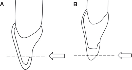
Fig. 2 (A) Specimen preparation design of Castelnuovo et al.,17 (B) Specimen preparation design of Schmidt et al.22

Fig. 3 Specimen preparation design of Chun et al.30 (A) labial veneer, (B) 3/4 veneer, enamel, (C) 3/4 veneer, dentin, (D) full veneer, enamel, (E) full veneer, dentin.
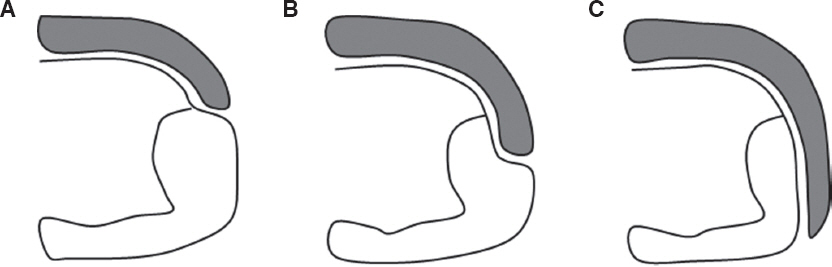
Fig. 4 The margin location of each veneer.41 (A) Short wrapping-the veneer was extended only to the facial margin of the preexisting interdental composite. (B) Medium wrapping-the veneer was extended into the bulk of the preexisting composite, penetrating 50% of the interdental area. (C) Long wrapping-the veneer covered the entire interdental area.
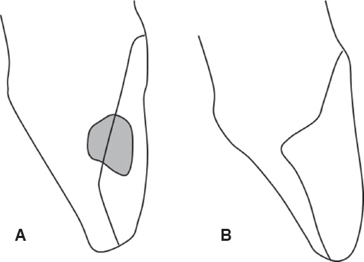
Fig. 5 The design of PLV preparation on tooth with class III composite filling (A), tooth with class IV cavity (B).42 PLV, porcelain laminate veneer.
Reference
-
References
1. Peumans M, De Munck J, Fieuws S, Lambrechts P, Vanherle G, Van Meerbeek B. A prospective tenyear clinical trial of porcelain veneers. J Adhes Dent. 2004; 6:65–76. PMID: 15119590.2. Burke FJ. Survival rates for porcelain laminate veneers with special reference to the effect of preparation in dentin: a literature review. J Esthet Restor Dent. 2012; 24:257–65. DOI: 10.1111/j.1708-8240.2012.00517.x. PMID: 22863131.3. Radz GM. Minimum thickness anterior porcelain restorations. Dent Clin North Am. 2011; 55:353–70. DOI: 10.1016/j.cden.2011.01.006. PMID: 21473998.4. Shetty A, Kaiwar A, Shubhashini N, Ashwini P, Naveen D, Adarsha M, Shetty M, Meena N. Survival rates of porcelain laminate restoration based on different incisal preparation designs: an analysis. J Conserv Dent. 2011; 14:10–5. DOI: 10.4103/0972-0707.80723. PMID: 21691498. PMCID: PMC3099105.5. Sadowsky SJ. An overview of treatment considerations for esthetic restorations: a review of the literature. J Prosthet Dent. 2006; 96:433–42. DOI: 10.1016/j.prosdent.2006.09.018. PMID: 17174661.6. Beier US, Kapferer I, Dumfahrt H. Clinical longterm evaluation and failure characteristics of 1,335 all-ceramic restorations. Int J Prosthodont. 2012; 25:70–8. PMID: 22259801.7. Beier US, Kapferer I, Burtscher D, Dumfahrt H. Clinical performance of porcelain laminate veneers for up to 20 years. Int J Prosthodont. 2012; 25:79–85. PMID: 22259802.8. Smales RJ, Etemadi S. Long term survival of porcelain laminate veneers using two preparation designs: a retrospective study. Int J Prosthodont. 2004; 17:323–6. PMID: 15237880.9. Rucker LM, Richter W, MacEntee M, Richardson A. Porcelain and resin veneers clinically evaluated 2- year results. J Am Dent Assoc. 1990; 121:594–6. DOI: 10.14219/jada.archive.1990.0225. PMID: 2229737.10. Meijering AC, Creugers NH, Roeters FJ, Mulder J. Survival of three types of veneer restorations in a clinical trial: a 2.5 year interim evaluation. J Dent. 1998; 26:563–8. DOI: 10.1016/S0300-5712(97)00032-8.11. Hui KK, Williams B, Davis EH, Holt RD. A comparative assessment of the strengths of porcelain veneers for incisor teeth dependent on their design characteristics. Br Dent J. 1991; 171:51–5. DOI: 10.1038/sj.bdj.4807602. PMID: 1873094.12. Highton R, Caputo AA, Mátyás J. A photoelastic study of stress on porcelain laminate preparations. J Prosthet Dent. 1987; 58:157–61. DOI: 10.1016/0022-3913(87)90168-5.13. Magne P, Douglas WH. Design optimization and evolution of bonded ceramics for anterior dentition: a finite-element analysis. Quintessence Int. 1999; 30:661–72. PMID: 10765850.14. Zarone F, Apicella D, Sorrentino R, Ferro V, Aversa R, Apicella A. Influence of tooth preparation design on the stress distribution in maxillary central incisors restored by means of alumina porcelain veneers: a 3D-finite element analysis. Dent Mater. 2005; 21:1178–88. DOI: 10.1016/j.dental.2005.02.014. PMID: 16098574.15. Magne P, Kwon KR, Belser UC, Hodges JS, Douglas WH. Crack propensity of porcelain laminate veneers: a simulated operatory evaluation. J Prosthet Dent. 1999; 81:327–34. DOI: 10.1016/S0022-3913(99)70277-5.16. Seymour KG, Cherukara GP, Samarawickrama DY. Stresses within porcelain veneers and the composite lute using different preparation designs. J Prosthodont. 2001; 10:16–21. DOI: 10.1111/j.1532-849X.2001.00016.x. PMID: 11406791.17. Castelnuovo J, Tjan AH, Phillips K, Nicholls JI, Kois JC. Fracture load and mode of failure of ceramic veneers with different preparations. J Prosthet Dent. 2000; 83:171–80. DOI: 10.1016/S0022-3913(00)80009-8.18. Hahn P, Gustav M, Hellwig E. An in vitro assessment of the strength of porcelain veneers dependent on tooth preparation. J Oral Rehabil. 2000; 27:1024–9. DOI: 10.1111/j.1365-2842.2000.00640.x. PMID: 11251771.19. Stappert CF, Ozden U, Gerds T, Strub JR. Longevity and failure load of ceramic veneers with different preparation designs after exposure to masticatory simulation. J Prosthet Dent. 2005; 94:132–9. DOI: 10.1016/j.prosdent.2005.05.023. PMID: 16046967.20. Zarone F, Epifania E, Leone G, Sorrentino R, Ferrari M. Dynamometric assessment of the mechanical resistance of porcelain veneers related to tooth preparation: a comparison between two techniques. J Prosthet Dent. 2006; 95:354–63. DOI: 10.1016/j.prosdent.2006.03.003. PMID: 16679130.21. Chaiyabutr Y, Phillips KM, Ma PS, Chitswe K. Comparison of load-fatigue testing of ceramic veneers with two different preparation designs. Int J Prosthodont. 2009; 22:573–5. PMID: 19918591.22. Schmidt KK, Chiayabutr Y, Phillips KM, Kois JC. Influence of preparation design and existing condition of tooth structure on load to failure of ceramic laminate veneers. J Prosthet Dent. 2011; 105:374–82. DOI: 10.1016/S0022-3913(11)60077-2.23. da Costa DC, Coutinho M, de Sousa AS, Ennes JP. A meta-analysis of the most indicated preparation design for porcelain laminate veneers. J Adhes Dent. 2013; 15:215–20. PMID: 23593640.24. Shetty A, Kaiwar A, Shubhashini N, Ashwini P, Naveen D, Adarsha M, Shetty M, Meena N. Survival rates of porcelain laminate restoration based on different incisal preparation designs: an analysis. J Conserv Dent. 2011; 14:10–5. DOI: 10.4103/0972-0707.80723. PMID: 21691498. PMCID: PMC3099105.25. Wall JG, Reisbick MH, Johnston WM. Incisal-edge strength of porcelain laminate veneers restoring mandibular incisors. Int J Prosthodont. 1992; 5:441–6. PMID: 1290573.26. Ako lu B, Gemalmaz D. Fracture resistance of ceramic veneers with different preparation designs. J Prosthodont. 2011; 20:380–4. DOI: 10.1111/j.1532-849X.2011.00728.x. PMID: 21631629.27. Rouse JS. Full veneer versus traditional veneer preparation: a discussion of interproximal extension. J Prosthet Dent. 1997; 78:545–9. DOI: 10.1016/S0022-3913(97)70003-9.28. Rosenthal L. Diastema closure utilizing porcelain veneers - simple and advanced. Dent Econ. 1994; 84:63–4. PMID: 7995439.29. Knight LD. Use of porcelain for treating a maxillary central diastema. Gen Dent. 1992; 40:498–9. PMID: 1298674.30. Chun YH, Raffelt C, Pfeiffer H, Bizhang M, Saul G, Blunck U, Roulet JF. Restoring strength of incisors with veneers and full ceramic crowns. J Adhes Dent. 2010; 12:45–54. PMID: 20155230.31. Guess PC, Stappert CF. Midterm results of a 5-year prospective clinical investigation of extended ceramic veneers. Dent Mater. 2008; 24:804–13. DOI: 10.1016/j.dental.2007.09.009. PMID: 18006051.32. Troedson M, Dérand T. Effect of margin design, cement polymerization, and angle of loading on stress in porcelain veneers. J Prosthet Dent. 1999; 82:518–24. DOI: 10.1016/S0022-3913(99)70049-1.33. Pahlevan A, Mirzaee M, Yassine E, Ranjbar OmraOmrany L, Hasani Tabatabaee M, Kermanshah H, Arami S, Abbasi M. Enamel thickness after preparation of tooth for porcelain laminate. J Dent (Tehran). 2014; 11:428–32.34. Rosenstiel SF, Land MF, Fujimoto J. Contemporary fixed prosthodontics. 4th ed. New York: Mosby;2006. p. 330–1.35. Burke FJ. Survival rates for porcelain laminate veneers with special reference to the effect of preparation in dentin: a literature review. J Esthet Restor Dent. 2012; 24:257–65. DOI: 10.1111/j.1708-8240.2012.00517.x. PMID: 22863131.36. Friedman MJ. A 15-year review of porcelain veneer failure -a clinician’s observations. Compend Contin Educ Dent. 1998; 19:625–8. PMID: 9693518.37. Peumans M, De Munck J, Fieuws S, Lambrechts P, Vanherle G, Van Meerbeek B. A prospective ten-year clinical trial of porcelain veneers. J Adhes Dent. 2004; 6:65–76. PMID: 15119590.38. Magne P, Douglas WH. Porcelain veneers: dentin bonding optimization and biomimetic recovery of the crown. Int J Prosthodont. 1999; 12:111–21. PMID: 10371912.39. Magne P, Kim TH, Cascione D, Donovan TE. Immediate dentin sealing improves bond strength of indirect restorations. J Prosthet Dent. 2005; 94:5119. DOI: 10.1016/j.prosdent.2005.10.010. PMID: 16316797.40. Magne P, So WS, Cascione D. Immediate dentin sealing supports delayed restoration placement. J Prosthet Dent. 2007; 98:166–74. DOI: 10.1016/S0022-3913(07)60052-3.41. Magne P, Douglas WH. Interdental design of porcelain veneers in the presence of composite fillings: finite element analysis of composite shrinkage and thermal stresses. Int J Prosthodont. 2000; 13:117–24. PMID: 11203619.42. Sadighpour L, Geramipanah F, Allahyari S, Fallahi Sichani B, Kharazi Fard MJ. In vitro evaluation of the fracture resistance and microleakage of porcelain laminate veneers bonded to teeth with composite fillings after cyclic loading. J Adv Prosthodont. 2014; 6:278–84. DOI: 10.4047/jap.2014.6.4.278. PMID: 25177471. PMCID: PMC4146728.43. Gresnigt MM, Ozcan M, Kalk W, Galhano G. Effect of static and cyclic loading on ceramic laminate veneers adhered to teeth with and without aged composite restorations. J Adhes Dent. 2011; 13:56977.44. Meijering AC, Creugers NH, Roeters FJ, Mulder J. Survival of three types of veneer restorations in a clinical trial: a 2.5-year interim evaluation. J Dent. 1998; 26:563–8. DOI: 10.1016/S0300-5712(97)00032-8.45. Shaini FU, Shortall AC, Marquis PM. Clinical performance of porcelain laminate veneers. A retrospective evaluation over a period of 6.5 years. J Oral Rehabil. 1997; 24:553–9. DOI: 10.1111/j.1365-2842.1997.tb00373.x. PMID: 9291247.46. Magne P, Douglas WH. Cumulative effects of successive restorative procedures on anterior crown flexure: intact versus veneered incisors. Quintessence Int. 2000; 31:5–18. PMID: 11203907.47. D’Arcangelo C, De Angelis F, Vadini M, Zazzeroni S, Ciampoli C, D’Amario M. In vitro fracture resistance and deflection of pulpless teeth restored with fiber posts and prepared for veneers. J Endod. 2008; 34:838–41. DOI: 10.1016/j.joen.2008.03.026. PMID: 18570991.48. D’Arcangelo C, De Angelis F, Vadini M, D’Amario M, Caputi S. Fracture resistance and deflection of pulpless anterior teeth restored with composite or porcelain veneers. J Endod. 2010; 36:153–6. DOI: 10.1016/j.joen.2009.09.036. PMID: 20003956.
- Full Text Links
-
- Actions
-
Cited
- CITED
-
- Close
- Share
-
- Similar articles
-
- Volume difference in upper central incisor preparation according to the changes of restorative design and marginal location
- THREE-DIMENSIONAL FINITE ELEMENT ANALYSIS OF STRESS DISTRIBUTION IN PORCELAIN LAMINATE VENEERS WITH VARIOUS AMOUNTS OF INCISAL COVERAGE AND TYPES OF INCISAL FINISH LINE UNDER TWO LOADING CONDITIONS
- The effect of the amount of interdental spacing on the stress distribution in maxillary central incisors restored with porcelain laminate veneer and composite resin: A 3D-finite element analysis
- Esthetic improvement in the patient with one missing maxillary central incisor restored with porcelain laminate veneers
- Influencing factors on the final color of laminate veneer restorations with various IPS Empress Esthetic(R) ingots
