J Adv Prosthodont.
2010 Sep;2(3):77-80. 10.4047/jap.2010.2.3.77.
Esthetic improvement in the patient with one missing maxillary central incisor restored with porcelain laminate veneers
- Affiliations
-
- 1Department of Prosthodontics and Dental research Institute, School of Dentistry, Seoul National University, Seoul, Korea. proshan@snu.ac.kr
- KMID: 2118110
- DOI: http://doi.org/10.4047/jap.2010.2.3.77
Abstract
- This article describes esthetic improvement in a patient with a missing maxillary left central incisor. Space analysis of the anterior dentition showed that minor tooth rearrangement was needed. Optimal space distribution for restorations was attained by orthodontic treatment. Through transforming tooth shape with porcelain laminate veneers, the maxillary left lateral incisor was transformed into central incisor and the maxillary left canine into a lateral incisor. The maxillary right central incisor was also restored for esthetic improvement. In a case of changing a tooth shape with porcelain laminate veneers, pre-treatment evaluation, space analysis and diagnostic wax-up are important factors.
Figure
-
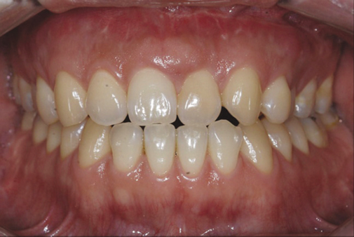
Fig. 1 Preoperative view.

Fig. 2 Minor tooth rearrangement was completed. The space of maxillary left lateral incisor became equal to that of maxillary right central incisor.

Fig. 3 Diagnostic wax-up. The maxillary left lateral incisor and maxillary left canine were transformed into a central incisor and lateral incisor.

Fig. 4 Frontal view of the tooth preparations.
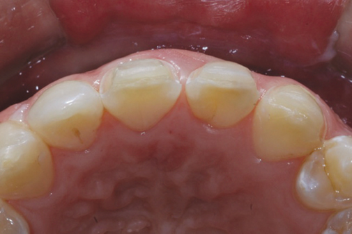
Fig. 5 Occlusal view of the tooth preparations.

Fig. 6 Porcelain laminate veneers were checked on the master cast for esthetics and adaptation.

Fig. 7 Frontal view of cemented restorations (1 month after delivery).
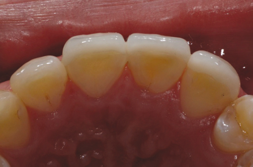
Fig. 8 Occlusal view of cemented restorations (1 month after delivery).
Reference
-
1. Claman L, Alfaro MA, Mercado A. An interdisciplinary approach for improved esthetic results in the anterior maxilla. J Prosthet Dent. 2003. 89:1–5.2. Aristidis GA, Dimitra B. Five-year clinical performance of porcelain laminate veneers. Quintessence Int. 2002. 33:185–189.3. Peumans M, De Munck J, Fieuws S, Lambrechts P, Vanherle G, Van Meerbeek B. A prospective ten-year clinical trial of porcelain veneers. J Adhes Dent. 2004. 6:65–76.4. Wiedhahn K, Kerschbaum T, Fasbinder DF. Clinical long-term results with 617 Cerec veneers: a nine-year report. Int J Comput Dent. 2005. 8:233–246.5. Fradeani M, Redemagni M, Corrado M. Porcelain laminate veneers: 6- to 12-year clinical evaluation--a retrospective study. Int J Periodontics Restorative Dent. 2005. 25:9–17.6. Swift EJ Jr, Friedman MJ. Critical appraisal: porcelain veneer outcomes, part II. J Esthet Restor Dent. 2006. 18:110–113.7. Magne P, Magne M, Belser UC. Adhesive restorations, centric relation, and the Dahl principle: minimally invasive approaches to localized anterior tooth erosion. Eur J Esthet Dent. 2007. 2:260–273.8. Griffin JD Jr. Correction of congenitally missing lateral incisors with porcelain veneers. Pract Proced Aesthet Dent. 2006. 18:475–480.9. Albakry M, Guazzato M, Swain MV. Biaxial flexural strength, elastic moduli, and x-ray diffraction characterization of three pressable all-ceramic materials. J Prosthet Dent. 2003. 89:374–380.
- Full Text Links
-
- Actions
-
Cited
- CITED
-
- Close
- Share
-
- Similar articles
-
- Esthetic restorations of maxillary anterior teeth with orthodontic treatment and porcelain laminate veneers: a case report
- The effect of the amount of interdental spacing on the stress distribution in maxillary central incisors restored with porcelain laminate veneer and composite resin: A 3D-finite element analysis
- In vitro evaluation of the fracture resistance and microleakage of porcelain laminate veneers bonded to teeth with composite fillings after cyclic loading
- Influencing factors on the final color of laminate veneer restorations with various IPS Empress Esthetic(R) ingots
- Anterior esthetic restoration using DSD (digital smile design) for a patient with congenital missing tooth of maxillary central incisor
