J Adv Prosthodont.
2014 Aug;6(4):278-284. 10.4047/jap.2014.6.4.278.
In vitro evaluation of the fracture resistance and microleakage of porcelain laminate veneers bonded to teeth with composite fillings after cyclic loading
- Affiliations
-
- 1Dental Research Center, Department of Prosthodontics, School of Dentistry, Tehran University of Medical Sciences, Tehran, Iran. somayeh.al@gmail.com
- 2Dental Implant Research Center, Department of Prosthodontics, School of Dentistry, Tehran University of Medical Sciences, Tehran, Iran. Geramopa@tums.ac.ir
- 3Research Institute for Nuclear Medicine, Shariati Hospital, Tehran university of Medical Sciences, Tehran, Iran.
- 4Department of Epidemiology, School of Public Health, Tehran University of Medical Sciences, Tehran, Iran.
- KMID: 1974847
- DOI: http://doi.org/10.4047/jap.2014.6.4.278
Abstract
- PURPOSE
There is insufficient data regarding the durability of porcelain laminate veneers bonded to existing composite fillings. The aim of the present study was to evaluate the fracture resistance and microleakage of porcelain laminate veneers bonded to teeth with existing composite fillings.
MATERIALS AND METHODS
Thirty maxillary central incisors were divided into three groups (for each group, n=10): intact teeth (NP), teeth with class III composite fillings (C3) and teeth with class IV cavities (C4). Porcelain laminate veneers were made using IPS-Empress ceramic and bonded with Panavia F2 resin cement. The microleakage of all of the specimens was tested before and after cyclic loading (1 x 10(6) cycles, 1.2 Hz). The fracture resistance values (N) were measured using a universal testing machine, and the mode of failure was also examined. The statistical analyses were performed using one-way ANOVA and Tukey post hoc tests (alpha=0.05).
RESULTS
There was a significant difference in the mean microleakage of group C4 compared with group NT (P=.013). There was no significant difference in the fracture loads among the groups.
CONCLUSION
The microleakage and failure loads of porcelain laminate veneers bonded to intact teeth and teeth with standard class III composite fillings were not significantly different.
Figure
-

Fig. 1 The design of PLV preparation on a tooth with class III composite filling. Blue lines demonstrates the position of inter proximal margin.

Fig. 2 The design of PLV preparation on a tooth with class IV cavity. The blue line demonstrates the position of interproximal margin.

Fig. 3 Specimens were painted by nail varnish to avoid the contamination with radioisotope.

Fig. 4 Teeth were mounted for the cyclic loading. The contact point was adjusted to the cingulum.
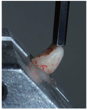
Fig. 5 The failure load was applied on the incisal surface.
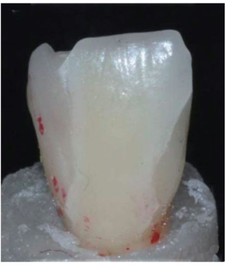
Fig. 6 An example of cohesive failure in laminates.
Reference
-
1. Calamia JR, Calamia CS. Porcelain laminate veneers: reasons for 25 years of success. Dent Clin North Am. 2007; 51:399–417.2. Peumans M, Van Meerbeek B, Lambrechts P, Vanherle G. Porcelain veneers: a review of the literature. J Dent. 2000; 28:163–177.3. Peumans M, De Munck J, Fieuws S, Lambrechts P, Vanherle G, Van Meerbeek B. A prospective ten-year clinical trial of porcelain veneers. J Adhes Dent. 2004; 6:65–76.4. Beier US, Kapferer I, Burtscher D, Dumfahrt H. Clinical performance of porcelain laminate veneers for up to 20 years. Int J Prosthodont. 2012; 25:79–85.5. Burke FJ. Survival rates for porcelain laminate veneers with special reference to the effect of preparation in dentin: a literature review. J Esthet Restor Dent. 2012; 24:257–265.6. Gresnigt MM, Ozcan M, Kalk W, Galhano G. Effect of static and cyclic loading on ceramic laminate veneers adhered to teeth with and without aged composite restorations. J Adhes Dent. 2011; 13:569–577.7. Guess PC, Stappert CF. Midterm results of a 5-year prospective clinical investigation of extended ceramic veneers. Dent Mater. 2008; 24:804–813.8. Chun YH, Raffelt C, Pfeiffer H, Bizhang M, Saul G, Blunck U, Roulet JF. Restoring strength of incisors with veneers and full ceramic crowns. J Adhes Dent. 2010; 12:45–54.9. Dennison JB, Sarrett DC. Prediction and diagnosis of clinical outcomes affecting restoration margins. J Oral Rehabil. 2012; 39:301–318.10. Magne P, Kwon KR, Belser UC, Hodges JS, Douglas WH. Crack propensity of porcelain laminate veneers: A simulated operatory evaluation. J Prosthet Dent. 1999; 81:327–334.11. Kuper NK, Opdam NJ, Bronkhorst EM, Ruben JL, Huysmans MC. Hydrodynamic flow through loading and in vitro secondary caries development. J Dent Res. 2013; 92:383–387.12. Gresnigt MM, Kalk W, Özcan M. Clinical longevity of ceramic laminate veneers bonded to teeth with and without existing composite restorations up to 40 months. Clin Oral Investig. 2013; 17:823–832.13. Summitt JB. Fundamentals of operative dentistry: A contemporary approach. 3rd ed. Chicago: Quintessence Pub;2006. p. 472.14. Behr M, Rosentritt M, Latzel D, Kreisler T. Comparison of three types of fiber-reinforced composite molar crowns on their fracture resistance and marginal adaptation. J Dent. 2001; 29:187–196.15. Stappert CF, Ozden U, Gerds T, Strub JR. Longevity and failure load of ceramic veneers with different preparation designs after exposure to masticatory simulation. J Prosthet Dent. 2005; 94:132–139.16. Öztürk E, Bolay Ş, Hickel R, Ilie N. Shear bond strength of porcelain laminate veneers to enamel, dentine and enamel-dentine complex bonded with different adhesive luting systems. J Dent. 2013; 41:97–105.17. Bayne SC. Dental restorations for oral rehabilitation - testing of laboratory properties versus clinical performance for clinical decision making. J Oral Rehabil. 2007; 34:921–932.18. Rueggeberg FA. Substrate for adhesion testing to tooth structure - review of the literature. Dent Mater. 1991; 7:2–10.19. Ozcan M, Mese A. Fracture strength of indirect resin composite laminates to teeth with existing restorations: an evaluation of conditioning protocols. J Adhes Dent. 2009; 11:391–397.20. Turkaslan S, Tezvergil-Mutluay A, Bagis B, Shinya A, Vallittu PK, Lassila LV. Effect of intermediate fiber layer on the fracture load and failure mode of maxillary incisors restored with laminate veneers. Dent Mater J. 2008; 27:61–68.21. Saraç YS, Başoğlu T, Ceylan GK, Saraç D, Yapici O. Effect of denture base surface pretreatment on microleakage of a silicone-based resilient liner. J Prosthet Dent. 2004; 92:283–287.22. Sarac D, Sarac YS, Basoglu T, Yapici O, Yuzbasioglu E. The evaluation of microleakage and bond strength of a silicone-based resilient liner following denture base surface pretreatment. J Prosthet Dent. 2006; 95:143–151.23. Geramipanah F, Rezaei SM, Sichani SF, Sichani BF, Sadighpour L. Microleakage of different post systems and a custom adapted fiber post. J Dent (Tehran). 2013; 10:94–102.24. Magne P, Douglas WH. Cumulative effects of successive restorative procedures on anterior crown flexure: intact versus veneered incisors. Quintessence Int. 2000; 31:5–18.25. Fradeani M, Redemagni M, Corrado M. Porcelain laminate veneers: 6- to 12-year clinical evaluation-a retrospective study. Int J Periodontics Restorative Dent. 2005; 25:9–17.26. Friedman MJ. Porcelain veneer restorations: a clinician's opinion about a disturbing trend. J Esthet Restor Dent. 2001; 13:318–327.27. Kasaz AC, Pena CE, de Alexandre RS, Viotti RG, Santana VB, Arrais CA, Giannini M, Reis AF. Effects of a peripheral enamel margin on the long-term bond strength and nanoleakage of composite/dentin interfaces produced by self-adhesive and conventional resin cements. J Adhes Dent. 2012; 14:251–263.
- Full Text Links
-
- Actions
-
Cited
- CITED
-
- Close
- Share
-
- Similar articles
-
- Esthetic restorations of maxillary anterior teeth with orthodontic treatment and porcelain laminate veneers: a case report
- Esthetic improvement in the patient with one missing maxillary central incisor restored with porcelain laminate veneers
- THREE-DIMENSIONAL FINITE ELEMENT ANALYSIS OF STRESS DISTRIBUTION IN PORCELAIN LAMINATE VENEERS WITH VARIOUS AMOUNTS OF INCISAL COVERAGE AND TYPES OF INCISAL FINISH LINE UNDER TWO LOADING CONDITIONS
- The effect of the amount of interdental spacing on the stress distribution in maxillary central incisors restored with porcelain laminate veneer and composite resin: A 3D-finite element analysis
- COMPARISON OF WEAR RESISTANCE AMONG RESIN DENTURE TEETH OPPOSING VARIOUS RESTORATIVE MATERIALS
