The effect of the amount of interdental spacing on the stress distribution in maxillary central incisors restored with porcelain laminate veneer and composite resin: A 3D-finite element analysis
- Affiliations
-
- 1Department of Conservative Dentistry, School of Dentistry, Seoul National University, Seoul, Korea. chobh@snu.ac.kr
- 2Mechanical Aerospace Engineering, Gyeongsang National University, Jinju, Korea.
- KMID: 2176275
- DOI: http://doi.org/10.5395/JKACD.2010.35.1.030
Abstract
- This study evaluated the influence of the type of restoration and the amount of interdental spacing on the stress distribution in maxillary central incisors restored by means of porcelain laminate veneers and direct composite resin restorations. Three-dimensional finite element models were fabricated to represent different types of restorations. Four clinical situations were considered. Type I, closing diastema using composite resin. Labial border of composite resin was extended just enough to cover the interdental space; Type II, closing diastema using composite resin without reduction of labial surface. Labial border of composite resin was extended distally to cover the half of the total labial surface; Type III, closing diastema using composite resin with reduction of labial surface. Labial border of the preparation and restored composite resin was extended distally two-thirds of the total labial surface; Type IV, closing diastema using porcelain laminate veneer with a feathered-edge preparation technique. Four different interdental spaces (1.0, 2.0, 3.0, 4.0 mm) were applied for each type of restorations. For all types of restoration, adding the width of free extension of the porcelain laminate veneer and composite resin increased the stress occurred at the bonding layer. The maximum stress values observed at the bonding layer of Type IV were higher than that of Type I, II and III. However, the increasing rate of maximum stress value of Type IV was lower than that of Type I, II and III.
Keyword
Figure
-

Figure 1 Finite element models of each type of restorations. a. Type I, b. Type II, c. Type III, d. Type IV.
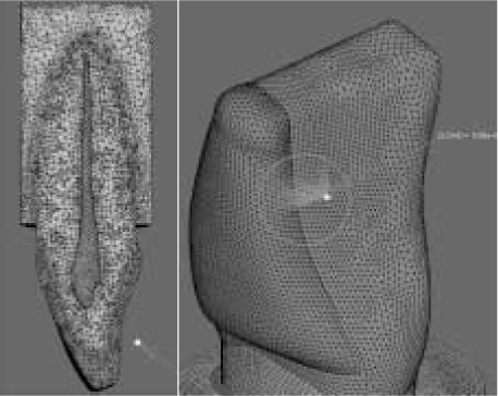
Figure 2 Load angulation. 50 N of load was applied at 125° angles (tearing force) with the tooth's longitudinal axis at the palatal surface of the crown.

Figure 3 von Mises stress distribution patterns of each type of restoration. These figures represent stress distribution at the tooth side of bonding layer. a. Type I with interdental space of 2.0 mm. b. Type II with interdental space of 2.0 mm. c. Type III with interdental space of 2.0 mm. d. Type IV with interdental space of 2.0 mm.
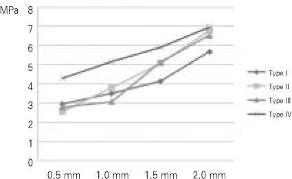
Figure 4 Line graphs of maximum von Mises stress values at the cervical area of the tooth side of bonding layer. Horizontal axis means interdental space. Vertical axis means von Mises stress values (MPa).
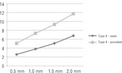
Figure 5 Maximum von Mises stress values in Type II restoration which was restored with porcelain laminate veneer instead of composite resin.
Reference
-
1. Heymann HO, Hershey HG. Use of composite resin for restorative and orthodontic correction of anterior interdental spacing. J Prosthet Dent. 1985. 53(6):766–771.
Article2. Lenhard M. Closing diastemas with resin composite restorations. Eur J Esthet Dent. 2008. 3(3):258–268.3. Pensler AV. Multiple-diastema porcelain laminate veneers: a case study. Compendium. 1993. 14(11):1470–1478.4. Nash RW. Closing a large central diastema using a pressed ceramic. Dent Today. 2003. 22(11):62–65.5. Aherne T. Treatment of maxillary anterior diastema using resin-bonded porcelain crown restorations. Pract Proced Aesthet Dent. 2001. 13(6):443–445.6. Tanaka OM, Furquim BD, Pascotto RC, Ribeiro GL, Bósio JA, Maruo H. The dilemma of the open gingival embrasure between maxillary central incisors. J Contemp Dent Pract. 2008. 9(6):92–98.
Article7. Dumfahrt H, Schaffer H. Porcelain laminate veneers. A retrospective evaluation after 1 to 10 years of service. Part II. Clinical results. Int J Prosthodont. 2000. 13:9–18.8. Peumans M, De Munck J, Fieuws S, Lambrechts P, Vanherle G, Van Meerbeek B. A prospective ten-year clinical trial of porcelain veneers. J Adhes Dent. 2004. 6:65–76.9. Burke FJ, Lucarotti PS. Ten-year outcome of porcelain laminate veneers placed within the general dental services in England and Wales. J Dent. 2009. 37:31–38.
Article10. Shaini FJ, Shortall AC, Marquis PM. Clinical performance of porcelain laminate veneers. A retrospective evaluation over a period of 6˙5 years. J Oral Rehabil. 1997. 24:553–559.
Article11. Dunne SM, Millar BJ. A longitudinal study of the clinical performance of porcelain veneers. Br Dent J. 1993. 175:317–321.
Article12. Hui KK, Williams B, Davis EH, Holt RD. A comparative assessment on the strengths of porcelain veneers for incisor teeth dependent on their design characteristics. Br Dent J. 1991. 171:51–55.
Article13. Highton R, Caputo AA, Mátyás J. A photoelastic study of stresses on porcelain laminate preparations. J Prosthet Dent. 1987. 58:157–161.
Article14. Hahn P, Gustav M, Hellwig E. An in vitro assessment of the strength of porcelain veneers dependent on tooth preparation. J Oral Rehabil. 2000. 27:1024–1029.
Article15. Morin DL, Douglas WH, Cross M, DeLong R. Biophysical stress analysis of restored teeth: experimental strain measurement. Dent Mater. 1988. 4:41–48.
Article16. Karl M, Dickinson A, Holst S, Holst A. Biomechanical methods applied in dentistry: a comparative overview of photoelastic examinations, strain gauge measurements, finite element analysis and three-dimensional deformation analysis. Eur J Prosthodont Restor Dent. 2009. 17(2):50–57.17. Reeh ES, Ross GK. Tooth stiffness with composite veneers: a strain gauge and finite element evaluation. Dent Mater. 1994. 10:247–252.
Article18. Morin DL, Cross M, Voller VR, Douglas WH, DeLong R. Biophysical stress analysis of restored teeth: modelling and analysis. Dent Mater. 1988. 4:77–84.
Article19. Hutton DV. Fundamentals of finite element analysis. 2006. 4:77–84.20. Cattaneo PM, Dalstra M, Melsen B. The finite element method: a tool to study orthodontic tooth movement. J Dent Res. 2005. 84:428–433.
Article21. Magne P, Belser UC. Rationalization of shape and related stress distribution in posterior teeth: a finite element study using nonlinear contact analysis. Int J Periodontics Restorative Dent. 2002. 22:425–433.22. Dejak B, Mlotkowski A, Romanowicz M. Finite element analysis of mechanism of cervical lesion formation in simulated molars during mastication and parafunction. J Prosthet Dent. 2005. 94:520–529.
Article23. Boccaccio A, Lamberti L, Pappalettere C, Cozzani M, Siciliani G. Comparison of different orthodontic devices for mandibular symphyseal distraction osteogenesis: A finite element study. Am J Orthod Dentofacial Orthop. 2008. 134:260–269.
Article24. Ona M, Wakabayashi N. Influence of alveolar support on stress in periodontal structures. J Dent Res. 2006. 85:1087–1091.
Article25. Versluis A, Tantbirojn D, Douglas WH. Do dental composite always shrink toward the light. J Dent Res. 1998. 77(6):1435–1445.26. Chun HJ, Shin HS, Han CH, Lee SH. Influence of implant abutment type on stress distribution in bone under various loading conditions using finite element analysis. Int J Oral Maxillofac Implants. 2006. 21:195–202.27. Hübsch PF, Middleton J, Knox J. A finite element analysis of the stress at the restoration-tooth interface, comparing inlays and bulk fillings. Biomaterials. 2000. 21:1015–1019.
Article28. Magne P, Douglas WH. Design optimization and evolution of bonded ceramics for the anterior dentition: A finite-element analysis. Quintessence Int. 1999. 30:661–672.29. Zarone F, Apicella D, Sorrentino R, Ferro V, Aversa R, Apicella A. Influence of tooth preparation design on the stress distribution in maxillary central incisors restored by means of alumina porcelain veneers: A 3D-finite element analysis. Dent Mater. 2005. 21:1178–1188.
Article30. Seymour KG, Cherukara GP, Samarawickrama DY. Stress within porcelain veneers and the composite lute using different preparation designs. J Prosthodont. 2001. 10:16–21.
Article31. Troedson M, Derand T. Shear stresses in the adhesive layer under porcelain veneers: A finite element method study. Acta Odontol Scand. 1998. 56:257–262.
Article32. Troedson M, Derand T. Effect of margin design, cement polymerization, and angle of loading on stress in porcelain veneers. J Prosthet Dent. 1999. 82:518–524.
Article33. Chander NG, Padmanabhan TV. Finite element stress analysis of diastema closure with ceramic laminate veneers. J Prosthodont. 2009. 18(7):577–581.
Article34. Dejak B, Mlotkowski A. Three-dimensional finite element analysis of strength and adhesion of composite resin versus ceramic inlays in molars. J Prosthet Dent. 2008. 99(2):131–140.
Article35. Anusavice K. Phillips' Science of Dental Materials. 1996. 10th ed. Philadelphia: Saunders;590–595.36. Baratieri LN. Direct adhesive restoration on fractured anterior teeth. 2000. 265–312.37. Baratieri LN. Composite Restorations in Anterior Teeth: Fundamentals and Possibilities. 2005. Sao Paulo: Quintessence books..38. Aschheim KW, Dale BG. Esthetic dentistry: a clinical approach to techniques and materials. 2001. 2nd ed. St. Louis: Mosby Inc.;151–155.39. Jordan RE, Suzuki M, Senda A. A clinical evaluation of porcelain laminate veneers: a four year recall report. J Esthet Dent. 1989. 1:126–137.40. Graber TM. Normal occlusion. Orthodontics, principles and practice. 1972. 3rd ed.WB Saunders Co..41. Carlsson GE. Bite force and chewing efficiency. Front Oral Physiol. 1974. 1:265–292.
Article42. Ausiello P, Rengo S, Davidson CL, Watts DC. Stress distributions in adhesively cemented ceramic and resin-composite Class II inlay restorations: a 3D-FEA study. Dent Mater. 2004. 20:862–872.
Article43. Craig RG. Compressive Properties of Enamel, Dental Cements, and Gold. J Dent Res. 1961. 40:936–945.
Article44. Craig RG. Restorative dental materials. 1985. St. Louis., MO: The C.V. Mosby Co.;–. .45. Sano H, Ciucchi B, Matthews WG, Pashley DH. Tensile properties of mineralized and demineralized human and bovine dentin. J Dent Res. 1994. 73:1205–1211.
Article46. Farah JW, Craig RG, Meroueh KA. Finite element analysis of three- and four-unit bridges. J Oral Rehabil. 1989. 16:603–611.
Article47. Lin CP. Structure-property-function relationships in the dentin-enamel complex and tooth-restoration interface(dissertation). 1993. Minneapolis, MN: University of Minnesota.48. Magne P, Perakis N, Belser UC, Krejci I. Stress distribution of inlay-anchored adhesive fixed partial dentures: a finite element analysis of influence of restorative materials and abutment preparation design. J Prosthet Dent. 2002. 87:516–527.49. Rees JS. Elastic modulus of the periodontal ligament. Biomaterials. 1997. 18:995–999.
Article50. Albakry M. Biaxial flexural strength, elastic moduli, and x-ray diffraction characterization of three pressable all-ceramic materials. J Prosthet Dent. 2003. 89:374–380.
Article51. Pegoretti A. Finite element analysis of a glass fibre reinforced composite endodontic post. Biomaterials. 2002. 23:2667–2682.
Article52. Farah JW, Graig RG. Finite element stress analysis of a restored axisymmetric first molar. J Dent Res. 1974. 53:859–866.
Article
- Full Text Links
-
- Actions
-
Cited
- CITED
-
- Close
- Share
-
- Similar articles
-
- THREE-DIMENSIONAL FINITE ELEMENT ANALYSIS OF STRESS DISTRIBUTION IN PORCELAIN LAMINATE VENEERS WITH VARIOUS AMOUNTS OF INCISAL COVERAGE AND TYPES OF INCISAL FINISH LINE UNDER TWO LOADING CONDITIONS
- Esthetic restorations of maxillary anterior teeth with orthodontic treatment and porcelain laminate veneers: a case report
- The effect of restorative materials on the stress distribution of class V composite resin restorations: a 3D finite element investigation
- The influence of combining composite resins with different elastic modulus on the stress distribution of Class V restoration: a three-dimensional finite element study
- Effect of the marginal position of prosthesis on stress distribution of teeth with abfraction lesion using finite element analysis
