Static and Dynamic Parameters in Patients With Degenerative Flat Back and Change After Corrective Fusion Surgery
- Affiliations
-
- 1Department of Medicine and Rehabilitation, Wooridul Spine Hospital, Seoul, Korea. j986802@hanmail.net
- 2Department of Neurosurgery, Wooridul Spine Hospital, Seoul, Korea.
- KMID: 2356654
- DOI: http://doi.org/10.5535/arm.2016.40.4.682
Abstract
OBJECTIVE
To evaluate characteristics of static and dynamic parameters in patients with degenerative flat back (DFB) and to compare degree of their improvement between successful and unsuccessful surgical outcome groups
METHODS
Forty-seven patients with DFB were included who took whole spine X-ray and three-dimensional motion analysis before and 6 months after corrective surgery. Forty-four subjects were selected as a control group. As static parameters, thoracic kyphosis (TK), thoracolumbar junction (TLJ), lumbar lordosis (LL), pelvic incidence (PI), sacral slope (SS), and pelvic tilt (PT) were measured. As dynamic parameters, maximal and minimal angle of pelvic tilt, lower limb joints, and thoracic and lumbar vertebrae column (dynamic TK and LL) in sagittal plane were obtained.
RESULTS
The DFB group showed smaller TK and larger LL, pelvic posterior tilt, hip flexion, knee flexion, and ankle dorsiflexion than the control group. Most of these parameters were significantly corrected by fusion surgery. Dynamic spinal parameters correlated with static spinal parameters. The successful group obtained significant improvement in maximal and minimal dynamic LL than the unsuccessful group.
CONCLUSION
The DFB group showed characteristic lower limb and spinal angles in dynamic and static parameters. Correlation between static and dynamic parameters was found in spinal segment. Dynamic LL was good predictor of successful surgical outcomes.
Keyword
MeSH Terms
Figure
-

Fig. 1 Spinal and pelvic parameters. (A) Spinal parameters including thoracic kyphosis (TK), thoracolumbar junction (TLJ), and lumbar lordosis (LL). (a) TK was measured from the T5 superior end plate to T12 inferior end plate. (b) TLJ was measured from the T10 superior end plate to L2 inferior end plate. (c) LL was measured from the T12 inferior end plate to S1 superior end plate by the Cobb method. (B) Pelvic parameters including pelvic tilt (PT), pelvic incidence (PI), and sacral slope (SS). (a) PT is defined as the angle between a vertical line originating at the center of the bicoxofemoral axis and a line drawn between the same point and the middle of the superior end plate of S1. (b) PI is defined as the angle between the line perpendicular to the sacral plate and the line connecting the midpoint of the sacral plate to the bicoxofemoral axis. (c) SS is the angle between the S1 superior end plate and a horizontal line.
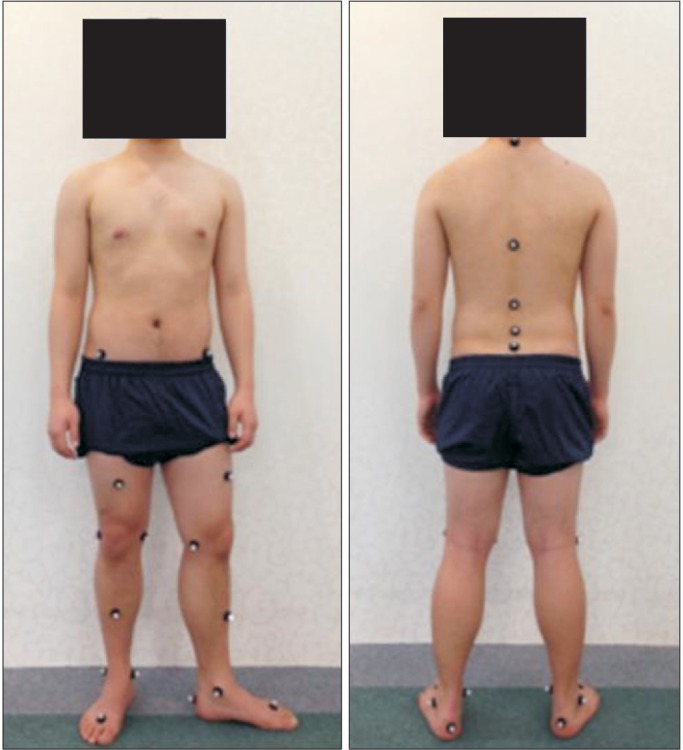
Fig. 2 Location of markers for motion analysis.
Reference
-
1. Lee JC, Cha JG, Kim Y, Kim YI, Shin BJ. Quantitative analysis of back muscle degeneration in the patients with the degenerative lumbar flat back using a digital image analysis: comparison with the normal controls. Spine (Phila Pa 1976). 2008; 33:318–325. PMID: 18303466.2. Takemitsu Y, Harada Y, Iwahara T, Miyamoto M, Miyatake Y. Lumbar degenerative kyphosis: clinical, radiological and epidemiological studies. Spine (Phila Pa 1976). 1988; 13:1317–1326. PMID: 2974629.3. Bae JS, Jang JS, Lee SH, Kim JU. Radiological analysis of lumbar degenerative kyphosis in relation to pelvic incidence. Spine J. 2012; 12:1045–1051. PMID: 23158969.
Article4. Kang CH, Shin MJ, Kim SM, Lee SH, Lee CS. MRI of paraspinal muscles in lumbar degenerative kyphosis patients and control patients with chronic low back pain. Clin Radiol. 2007; 62:479–486. PMID: 17398274.
Article5. Lee CS, Lee CK, Kim YT, Hong YM, Yoo JH. Dynamic sagittal imbalance of the spine in degenerative flat back: significance of pelvic tilt in surgical treatment. Spine (Phila Pa 1976). 2001; 26:2029–2035. PMID: 11547204.6. Sarwahi V, Boachie-Adjei O, Backus SI, Taira G. Characterization of gait function in patients with postsurgical sagittal (flatback) deformity: a prospective study of 21 patients. Spine (Phila Pa 1976). 2002; 27:2328–2337. PMID: 12438980.7. Barrey C, Roussouly P, Perrin G, Le Huec JC. Sagittal balance disorders in severe degenerative spine. Can we identify the compensatory mechanisms? Eur Spine J. 2011; 20(Suppl 5):626–633. PMID: 21796393.
Article8. Jang JS, Lee SH, Min JH, Maeng DH. Changes in sagittal alignment after restoration of lower lumbar lordosis in patients with degenerative flat back syndrome. J Neurosurg Spine. 2007; 7:387–392. PMID: 17933311.
Article9. Jang JS, Lee SH, Min JH, Maeng DH. Influence of lumbar lordosis restoration on thoracic curve and sagittal position in lumbar degenerative kyphosis patients. Spine (Phila Pa 1976). 2009; 34:280–284. PMID: 19179923.
Article10. Khodadadeh S, Eisenstein SM. Gait analysis of patients with low back pain before and after surgery. Spine (Phila Pa 1976). 1993; 18:1451–1455. PMID: 8235815.
Article11. McGinley JL, Baker R, Wolfe R, Morris ME. The reliability of three-dimensional kinematic gait measurements: a systematic review. Gait Posture. 2009; 29:360–369. PMID: 19013070.
Article12. Shum GL, Crosbie J, Lee RY. Three-dimensional kinetics of the lumbar spine and hips in low back pain patients during sit-to-stand and stand-to-sit. Spine (Phila Pa 1976). 2007; 32:E211–E219. PMID: 17414896.
Article13. Suda Y, Saitou M, Shibasaki K, Yamazaki N, Chiba K, Toyama Y. Gait analysis of patients with neurogenic intermittent claudication. Spine (Phila Pa 1976). 2002; 27:2509–2513. PMID: 12435983.
Article14. Kim WJ, Kang JW, Kang SI, Sung HI, Park KY, Park JG, et al. Factors affecting clinical results after corrective osteotomy for lumbar degenerative kyphosis. Asian Spine J. 2010; 4:7–14. PMID: 20622949.
Article15. Macnab I. Negative disc exploration: an analysis of the causes of nerve-root involvement in sixty-eight patients. J Bone Joint Surg Am. 1971; 53:891–903. PMID: 4326746.16. Ahn Y, Lee SH, Park WM, Lee HY, Shin SW, Kang HY. Percutaneous endoscopic lumbar discectomy for recurrent disc herniation: surgical technique, outcome, and prognostic factors of 43 consecutive cases. Spine (Phila Pa 1976). 2004; 29:E326–E332. PMID: 15303041.
Article17. Legaye J, Duval-Beaupère G, Hecquet J, Marty C. Pelvic incidence: a fundamental pelvic parameter for three-dimensional regulation of spinal sagittal curves. Eur Spine J. 1998; 7:99–103. PMID: 9629932.
Article
- Full Text Links
-
- Actions
-
Cited
- CITED
-
- Close
- Share
-
- Similar articles
-
- Which Criterion Is More Reliable for Selecting the Distal Fusion Level in Cases of Adolescent Idiopathic Scoliosis with Structural Thoracolumbar/Lumbar Curves: Static or Dynamic?
- The Result of Minimal Invasive Anterior Lumbar Interbody Fusion with Posterior Lumbar Interbody Fusion on Degenerative Lumbar Flat Back Disease
- Anterior Lumbar Interbody Fusion for Focal Type of Degenerative Flat Back: Preliminary Report
- A Surgical Treatment of Low-Grade Spondylolisthesis: A Comparative Study of in Situ Fusion and Fusion after Reduction
- Effects of Acute Low Back Pain on Postural Control
