Clin Endosc.
2016 Sep;49(5):404-407. 10.5946/ce.2016.100.
Raman Spectroscopy for the Endoscopic Diagnosis of Esophageal, Gastric, and Colonic Diseases
- Affiliations
-
- 1Division of Gastroenterology and Hepatology, National University Health System, Singapore. khek_yu_ho@nuhs.edu.sg
- KMID: 2356043
- DOI: http://doi.org/10.5946/ce.2016.100
Abstract
- Globally white-light endoscopy with biopsy sampling is the gold standard diagnostic modality for esophageal, gastric, and colonic pathologies. However, there is overwhelming evidence to highlight the deficiencies of an approach based predominantly on eyeball visualization. Biopsy sampling is also problematic due in part to excessive sampling and hence attendant cost. Various innovations are currently taking place in the endoscopic domain to aid operators in diagnosis forming. These include narrow band imaging which aims to enhance the surface anatomy and vasculature, and confocal laser endomicroscopy which provides real time histological information. However, both of these tools are limited by the skill of the operator and the extensive learning curve associated with their use. There is a gap therefore for a new form of technology that relies solely on an objective measure of disease and reduces the need for biopsy sampling. Raman spectroscopy (RS) is a potential platform that aims to satisfy these criteria. It enables a fingerprint capture of tissue in relation to the protein, DNA, and lipid content. This focused review highlights the strong potential for the use of RS during endoscopic gastroenterological examination.
Keyword
MeSH Terms
Figure
-
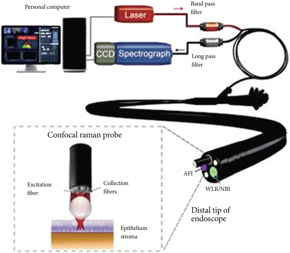
Fig. 1. Schematic diagram of the custom-built in vivo Raman spectroscopy system at National University Health System. Adapted from Bergholt et al. [1], with permission from Elsevier. CCD, charge coupled device; AFI, autofluorescence imaging; WLR, white-light reflectance; NBI, narrow band imaging.
Reference
-
1. Bergholt MS, Zheng W, Ho KY, et al. Fiberoptic confocal raman spectroscopy for real-time in vivo diagnosis of dysplasia in Barrett’s esophagus. Gastroenterology. 2014; 146:27–32.
Article2. Wang YW, Kang S, Khan A, Bao PQ, Liu JT. In vivo multiplexed molecular imaging of esophageal cancer via spectral endoscopy of topically applied SERS nanoparticles. Biomed Opt Express. 2015; 6:3714–3723.
Article3. Almond LM, Hutchings J, Lloyd G, et al. Endoscopic Raman spectroscopy enables objective diagnosis of dysplasia in Barrett’s esophagus. Gastrointest Endosc. 2014; 79:37–45.4. Almond LM, Hutchings J, Kendall C, et al. Assessment of a custom-built Raman spectroscopic probe for diagnosis of early oesophageal neoplasia. J Biomed Opt. 2012; 17:081421–081421.
Article5. Bergholt MS, Zheng W, Lin K, et al. In vivo diagnosis of esophageal cancer using image-guided Raman endoscopy and biomolecular modeling. Technol Cancer Res Treat. 2011; 10:103–112.
Article6. Wang J, Lin K, Zheng W, et al. Fiber-optic Raman spectroscopy for in vivo diagnosis of gastric dysplasia. Faraday Discuss. 2016; 187:377–392.
Article7. Lin K, Wang J, Zheng W, et al. Rapid fiber-optic Raman spectroscopy for real-time in vivo detection of gastric intestinal metaplasia during clinical gastroscopy. Cancer Prev Res (Phila). 2016; 9:476–483.
Article8. Luo S, Chen C, Mao H, Jin S. Discrimination of premalignant lesions and cancer tissues from normal gastric tissues using Raman spectroscopy. J Biomed Opt. 2013; 18:067004.
Article9. Bergholt MS, Zheng W, Ho KY, et al. Fiber-optic Raman spectroscopy probes gastric carcinogenesis in vivo at endoscopy. J Biophotonics. 2013; 6:49–59.10. Duraipandian S, Sylvest Bergholt M, Zheng W, et al. Real-time Raman spectroscopy for in vivo, online gastric cancer diagnosis during clinical endoscopic examination. J Biomed Opt. 2012; 17:081418.11. Kawabata T, Kikuchi H, Okazaki S, et al. Near-infrared multichannel Raman spectroscopy with a 1064 nm excitation wavelength for ex vivo diagnosis of gastric cancer. J Surg Res. 2011; 169:e137–e143.
Article12. Chen Y, Dai J, Zhou X, Liu Y, Zhang W, Peng G. Raman spectroscopy analysis of the biochemical characteristics of molecules associated with the malignant transformation of gastric mucosa. PLoS One. 2014; 9:e93906.
Article13. Bergholt MS, Lin K, Wang J, et al. Simultaneous fingerprint and high-wavenumber fiber-optic Raman spectroscopy enhances real-time in vivo diagnosis of adenomatous polyps during colonoscopy. J Biophotonics. 2016; 9:333–342.14. Bergholt MS, Zheng W, Lin K, et al. Characterizing variability of in vivo Raman spectroscopic properties of different anatomical sites of normal colorectal tissue towards cancer diagnosis at colonoscopy. Anal Chem. 2015; 87:960–966.15. Veenstra MA, Palyvoda O, Alahwal H, et al. Raman spectroscopy in the diagnosis of ulcerative colitis. Eur J Pediatr Surg. 2015; 25:56–59.
Article
- Full Text Links
-
- Actions
-
Cited
- CITED
-
- Close
- Share
-
- Similar articles
-
- Analysis of Normal and Cancer Tissue in the Stomach Using Raman Spectroscopy
- Application of Raman spectroscopy in breast cancer surgery
- Practical Approach to Endoscopic Management for Bleeding Gastric Varices
- Molecular Imaging for Theranostics in Gastroenterology: One Stone to Kill Two Birds
- Usefullness of Raman Spectroscopy in Differentiation between Cancer and Adjacent Normal Tissue of the Larynx
