Obstructive ileus caused by phlebosclerotic colitis
- Affiliations
-
- 1Department of Internal Medicine, Inje University Ilsan Paik Hospital, Goyang, Korea. jwkim@paik.ac.kr
- KMID: 2354956
- DOI: http://doi.org/10.5217/ir.2016.14.4.369
Abstract
- A 57-year-old man with chronic kidney disease and a history of using numerous herbal medications visited Inje University Ilsan Paik Hospital for abdominal pain and vomiting. An abdominal radiograph showed diffuse small bowel distension containing multiple air-fluid levels and extensive calcifications along the colon. Computed tomography showed colon wall thickening with diffuse calcification along the colonic mesenteric vein and colonic wall. Colonoscopy, performed without bowel preparation, showed bluish edematous mucosa from the transverse to the distal sigmoid colon, with multiple scar changes. At the mid transverse colon, a stricture was noted and the scope could not pass through. A biopsy of the stricture site revealed nonspecific changes. The patient was diagnosed with phlebosclerotic colitis. After the colonoscopy, the obstructive ileus spontaneously resolved, and the patient was discharged without an operation. Currently, after 2 months of follow-up, the patient has remained asymptomatic. Herein, we report the rare case of an obstructive ileus caused by phlebosclerotic colitis with a colon stricture.
MeSH Terms
Figure
-
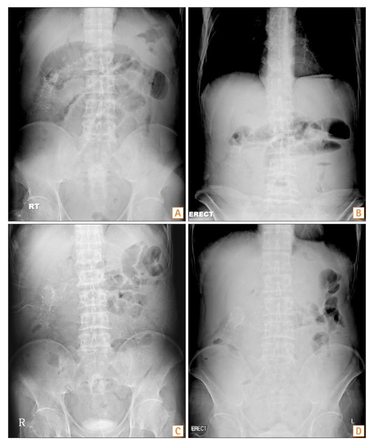
Fig. 1 Plain abdominal radiograph. Initial plain abdominal radiograph shows a diffuse small bowel ileus with multiple air-fluid levels and numerous calcification densities along the colon. (A) Supine, (B) Erect. Dramatically improved ileus, 1 day after the colonoscopy. (C) Supine, (D) Erect.
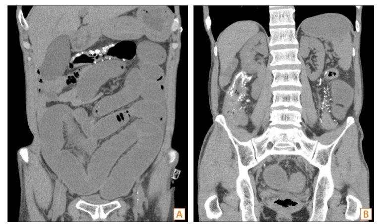
Fig. 2 Non-contrast-enhanced abdominal CT scan. (A) Diffuse fluid-filled distension of A B small bowel. (B) Colonic wall calcifications.
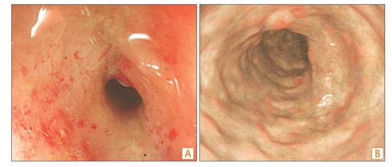
Fig. 3 Colonoscopy findings. (A) Pinpoint stricture in the transverse colon, scope passage was not possible. (B) Blue-grayish congestive change with diffuse scarring found in the transverse colon.
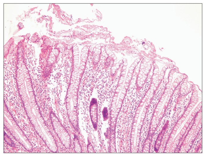
Fig. 4 Pathologic findings at the stricture site. The mucosa shows lymphoid cell infiltration and mild architectural distortion of crypts, which are suggestive of regenerative changes (H&E, ×100). No submucosal vessel was included in the specimen.
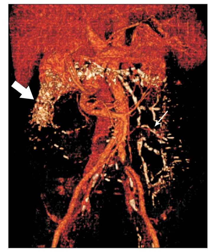
Fig. 5 Contrast-enhanced abdominal CT angiography. Abdominal CT scan shows extensive calcifications along the colonic wall (thick arrow) and mesenteric veins (thin arrow).
Reference
-
1. Iwashita A, Yao T, Schlemper RJ, et al. Mesenteric phlebosclerosis: a new disease entity causing ischemic colitis. Dis Colon Rectum. 2003; 46:209–220. PMID: 12576895.2. Chang KM. New histologic findings in idiopathic mesenteric phlebosclerosis: clues to its pathogenesis and etiology--probably ingested toxic agent-related. J Chin Med Assoc. 2007; 70:227–235. PMID: 17591581.
Article3. Song JH, Kim JI, Jung JH, et al. A case of phlebosclerotic colitis in a hemodialysis patient. Korean J Gastroenterol. 2012; 59:40–43. PMID: 22289953.
Article4. Kang HY, Noh R, Kim SM, Shin HD, Yun SY, Song IH. Phlebosclerotic colitis in a cirrhotic patient with portal hypertension: the first case in Korea. J Korean Med Sci. 2009; 24:1195–1199. PMID: 19949682.
Article5. Yao T, Iwashita A, Hoashi T, et al. Phlebosclerotic colitis: value of radiography in diagnosis--report of three cases. Radiology. 2000; 214:188–192. PMID: 10644121.
Article6. Hu P, Deng L. Phlebosclerotic colitis: three cases and literature review. Abdom Imaging. 2013; 38:1220–1224. PMID: 23589075.
Article7. Markos V, Kelly S, Yee WC, Davis JE, Cheifetz RE, Alsheikh A. Phlebosclerotic colitis: imaging findings of a rare entity. AJR Am J Roentgenol. 2005; 184:1584–1586. PMID: 15855120.
Article8. Hiramatsu K, Sakata H, Horita Y, et al. Mesenteric phlebosclerosis associated with long-term oral intake of geniposide, an ingredient of herbal medicine. Aliment Pharmacol Ther. 2012; 36:575–586. PMID: 22817400.
Article9. Choi JM, Lee KN, Kim HS, et al. Idiopathic phlebosclerotic colitis: a rare entity of chronic ischemic colitis. Korean J Gastroenterol. 2014; 63:183–186. PMID: 24651592.
Article10. Hirasaki S, Matsumura K. Development of phlebosclerotic colitis under treatment with Chinese herbal therapy. Intern Med. 2014; 53:1709–1710. PMID: 25088891.
Article11. Chang YY, Lin HH, Lin CC. Phlebosclerotic colitis presenting as intestinal obstruction. Clin Gastroenterol Hepatol. 2014; 12:e81–e82. DOI: 10.1016/j.cgh.2014.02.018. PMID: 24561098.
Article
- Full Text Links
-
- Actions
-
Cited
- CITED
-
- Close
- Share
-
- Similar articles
-
- A Case of Phlebosclerotic Colitis in a Hemodialysis Patient
- Phlebosclerotic Colitis in a Healthy Young Woman
- A Case of Phlebosclerotic Colitis in a Patient of Chronic Renal Failure
- Idiopathic Phlebosclerotic Colitis: A Rare Entity of Chronic Ischemic Colitis
- Phlebosclerotic Colitis in a Healthy Young Female with Long-term Herbal Medicine Use
