Phlebosclerotic Colitis in a Healthy Young Woman
- Affiliations
-
- 1Department of Internal Medicine, Pohang St. Mary's Hospital, Pohang, Korea. yyhhsung@hanmail.net
- KMID: 2148560
- DOI: http://doi.org/10.5946/ce.2015.48.5.447
Abstract
- Phlebosclerotic colitis is a rare disease of intestinal ischemia and differentiating it from the typical ischemic colitis. It is caused by venous obstruction due to colonic and mesenteric venous calcification. We report a 36-year-old woman presenting with intermittent abdominal pain. Initial radiologic findings showed multiple tortuous thread-like calcifications in the region of the right side of the colon and transverse colon on plain abdominal radiographs and computed tomography images. In the colonoscopy, edematous dark-bluish colonic mucosa, sclerotic colon wall, and multiple ulcers without clear boundaries were observed from the ascending colon to the transverse colon. In the sigmoid colon only showed the edematous dark-bluish colonic mucosa, sclerotic colon wall. On the basis of these findings, we diagnosed the patient as having phlebosclerotic colitis. We report a rare case of phlebosclerotic colitis in healthy young woman.
MeSH Terms
Figure
-
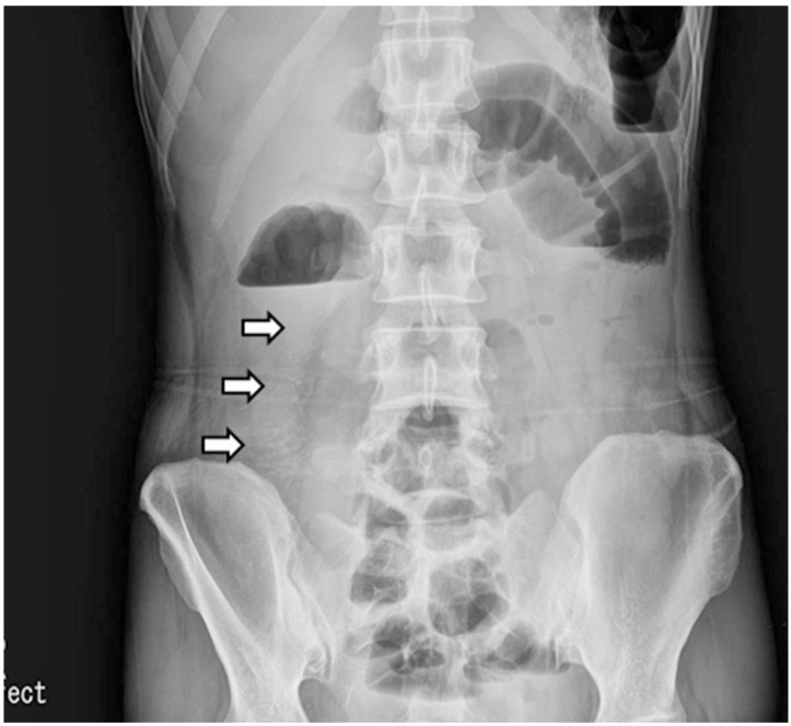
Fig. 1 Plain abdominal radiographic finding. It showed multiple linear calcifications in the right lower quadrant (white arrows).
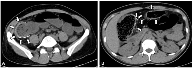
Fig. 2 (A, B) Abdominopelvic computed tomography finding. The enhanced computed tomography showed the thickening of the colonic wall with calcifications (white arrows).
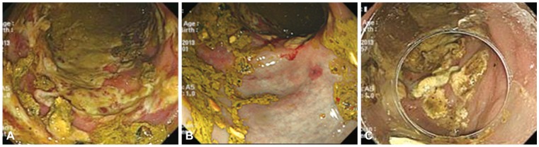
Fig. 3 Colonoscopic findings. (A, B) It noted dark blue colored edematous mucosa and ulcerations without clear boundaries from ascending colon to the sigmoid colon. (C) The follow-up colonoscopy. Improved fine ulcers were found in the ascending colon after 10 days of a discharge.
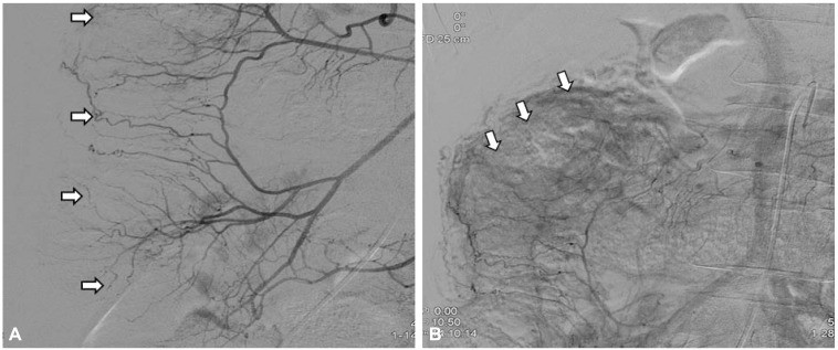
Fig. 4 Superior mesenteric angiography. (A) It indicated irregularity and tortuosity (arrows) of marginal arteries and vasa recta in right colic area at the arterial phase. (B) It indicated venous pooling (arrows) along the ascending colon at the delayed phase.
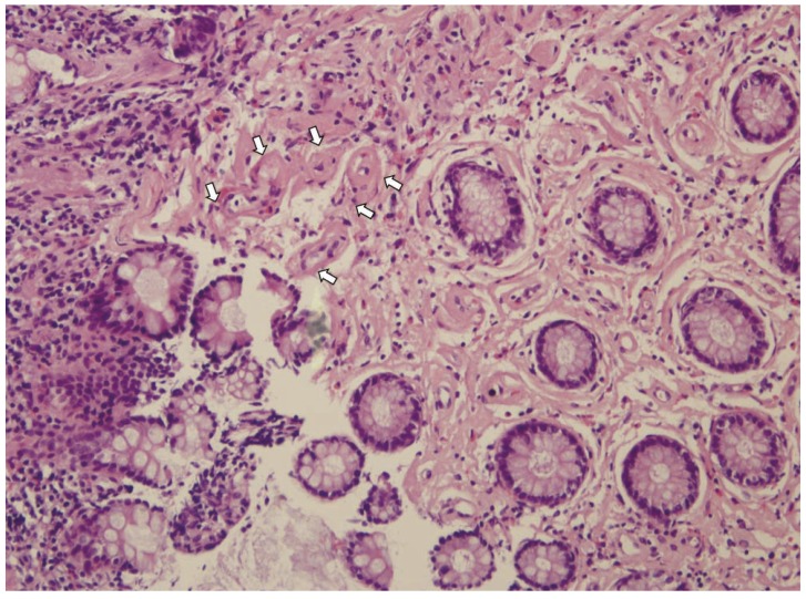
Fig. 5 Histologic finding. It shows tortuous veins (arrows) and fibrosis in vessel wall of ascending colonic mucosa (H&E stain, ×200).
Reference
-
1. Yao T, Iwashita A, Hoashi T, et al. Phlebosclerotic colitis: value of radiography in diagnosis: report of three cases. Radiology. 2000; 214:188–192. PMID: 10644121.2. Kusanagi M, Matsui O, Kawashima H, et al. Phlebosclerotic colitis: imaging-pathologic correlation. AJR Am J Roentgenol. 2005; 185:441–447. PMID: 16037518.
Article3. Kang HY, Noh R, Kim SM, Shin HD, Yun SY, Song IH. Phlebosclerotic colitis in a cirrhotic patient with portal hypertension: the first case in Korea. J Korean Med Sci. 2009; 24:1195–1199. PMID: 19949682.
Article4. Hu P, Deng L. Phlebosclerotic colitis: three cases and literature review. Abdom Imaging. 2013; 38:1220–1224. PMID: 23589075.
Article5. Yu SC, Kim JI, Lee SY, et al. A case of phlebosclerotic colitis in a patient of chronic renal failure. Korean J Gastrointest Endosc. 2009; 38:107–110.6. Song JH, Kim JI, Jung JH, et al. A case of phlebosclerotic colitis in a hemodialysis patient. Korean J Gastroenterol. 2012; 59:40–43. PMID: 22289953.
Article7. Choi JM, Lee KN, Kim HS, et al. Idiopathic phlebosclerotic colitis: a rare entity of chronic ischemic colitis. Korean J Gastroenterol. 2014; 63:183–186. PMID: 24651592.
Article8. Hur CJ, Kim EY, Oh JS, et al. A case of idiopathic mesenteric phlebosclerosis. Korean J Gastrointest Endosc. 2009; 38:352–355.9. Jung HG, Koh JW, Lee MY. A case of idiopathic mesenteric phlebosclerosis. Korean J Gastroenterol. 2008; 52:261–264. PMID: 19077529.10. Markos V, Kelly S, Yee WC, Davis JE, Cheifetz RE, Alsheikh A. Phlebosclerotic colitis: imaging findings of a rare entity. AJR Am J Roentgenol. 2005; 184:1584–1586. PMID: 15855120.
Article11. Maruyama Y, Watanabe F, Kanaoka S, et al. A case of phlebosclerotic ischemic colitis: a distinct entity. Endoscopy. 1997; 29:334. PMID: 9255548.
Article12. Chen MT, Yu SL, Yang TH. A case of phlebosclerotic colitis with involvement of the entire colon. Chang Gung Med J. 2010; 33:581–585. PMID: 20979710.13. Saito Y, Taniguchi M, Tagawa K, Ibukuro K, Mori M, Emura F. Phlebosclerotic colitis with deep circumferential ulceration: three-year endoscopic follow-up. Report of a case. Dis Colon Rectum. 2005; 48:2347–2351. PMID: 16258707.
Article14. Hozumi H, Hokari R, Shimizu M, et al. Phlebosclerotic colitis that was difficult to distinguish from collagenous colitis. Dig Endosc. 2014; 26:594–598. PMID: 23902595.
Article15. Kato T, Miyazaki K, Nakamura T, Tan KY, Chiba T, Konishi F. Perforated phlebosclerotic colitis: description of a case and review of this condition. Colorectal Dis. 2010; 12:149–151. PMID: 19175648.16. Tamamoto F, Ishizaki H, Maehara T. Phlebosclerotic colitis. Radiat Med. 2008; 26:164–167. PMID: 18683572.
Article17. Chang YY, Lin HH, Lin CC. Phlebosclerotic colitis presenting as intestinal obstruction. Clin Gastroenterol Hepatol. 2014; 12:e81–e82. PMID: 24561098.
Article18. Yu CJ, Wang HH, Chou JW, et al. Phlebosclerotic colitis with nonsurgical treatment. Int J Colorectal Dis. 2009; 24:1241–1242. PMID: 19390857.
Article19. Oshitani N, Matsumura Y, Kono M, et al. Asymptomatic chronic intestinal ischemia caused by idiopathic phlebosclerosis of mesenteric vein. Dig Dis Sci. 2002; 47:2711–2714. PMID: 12498290.20. Iwashita A, Yao T, Schlemper RJ, et al. Mesenteric phlebosclerosis: a new disease entity causing ischemic colitis. Dis Colon Rectum. 2003; 46:209–220. PMID: 12576895.
- Full Text Links
-
- Actions
-
Cited
- CITED
-
- Close
- Share
-
- Similar articles
-
- Phlebosclerotic Colitis in a Healthy Young Female with Long-term Herbal Medicine Use
- Idiopathic Phlebosclerotic Colitis: A Rare Entity of Chronic Ischemic Colitis
- A Case of Phlebosclerotic Colitis in a Hemodialysis Patient
- A Case of Phlebosclerotic Colitis in a Patient of Chronic Renal Failure
- Phlebosclerotic Colitis in a Cirrhotic Patient with Portal Hypertension: The First Case in Korea
