Enhancement of Optic Nerve in Leukemic Patients: Leukemic Infiltration of Optic Nerve versus Optic Neuritis
- Affiliations
-
- 1Department of Radiology, Seoul St. Mary's Hospital, College of Medicine, The Catholic University of Korea, Seoul, Korea. saim@catholic.ac.kr
- 2Department of Pediatrics, Seoul St. Mary's Hospital, College of Medicine, The Catholic University of Korea, Seoul, Korea.
- KMID: 2354797
- DOI: http://doi.org/10.13104/imri.2016.20.3.167
Abstract
- PURPOSE
To identify magnetic resonance imaging (MRI) findings of leukemic infiltration of optic nerve and optic neuritis in leukemic patients with emphasis of clinical findings as reference standard to differentiate them.
MATERIALS AND METHODS
MRI and clinical findings of 7 patients diagnosed as leukemic infiltration of optic nerve (n = 5) and optic neuritis (n = 2) in our institution between July 2006 and August 2015were reviewed retrospectively. In particular, MR imaging findings involved perineural enhancement and thickening of optic nerve and its degree, signal intensity, laterality (unilateral/bilateral), intraconal fat infiltration and its degree, and associated central nervous system abnormalities.
RESULTS
Of 5 cases of leukemic infiltration of optic nerve, 4 cases showed positive cerebrospinal fluid (CSF) study for leukemia relapse and 1 case was positive on bone marrow (BM) biopsy only. Moreover, of 5 leukemic infiltration of optic nerve, 2 cases showed the most specific MR findings for leukemic central nervous system involvement including 1 prominent leptomeningeal enhancement and 1 chloroma. However, other MR imaging findings of the patients with leukemic infiltration or optic neuritis such as thickening and perineural enhancement of optic nerves are overlapped.
CONCLUSION
Enhancement and thickening of optic nerve were overlapped MR findings in leukemic infiltration of optic nerve and optic neuritis. Our findings suggest that enhancing optic nerve thickening with associated central nervous system MR abnormality favors the diagnosis of leukemic infiltration of optic nerve, especially in patients with history of acute lymphoblastic leukemia. However, CSF and BM study were required for differentiation between leukemic infiltration of optic nerve and optic neuritis.
MeSH Terms
Figure
-
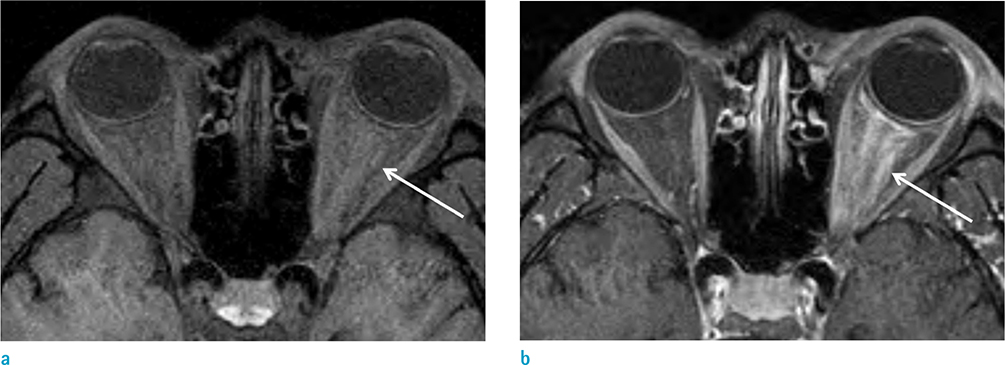
Fig. 1 Leukemic infiltration of left optic nerve in a 27-year-old man. He had a complaint of painful swelling of left eye, which occurred during chemotherapy after achieving second complete remission of acute myelogenous leukemia. (a, b) Axial fat-suppressed T1-weighted MR image (a) and contrast-enhanced, fat-suppressed, axial T1-weighted MR image (b) show diffuse thickening and perineural enhancement of left optic nerve with intraconal fat infiltration (arrows).
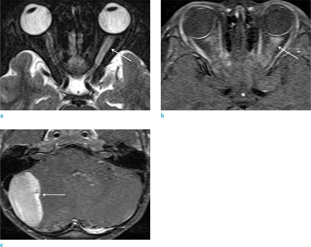
Fig. 2 Leukemic infiltration of left optic nerve in a 7-year-old boy. He complained of decreased visual acuity of left orbit and fever, which occurred during chemotherapy for acute lymphoblastic leukemia. (a) Fat-suppressed axial T2-weighted MR image shows thickening and high IS of left optic nerve (arrow). (b) Contrast-enhanced, fat-suppressed axial T1-weighted MR image shows diffuse thickening with perineural enhancement of left optic nerve with intraconal fat infiltration (arrow). (c) Contrast-enhanced axial T1-weighted MR image with fat-suppression of the brain demonstrates a chloroma arising from dura and extension to right cerebellar hemisphere (arrow).
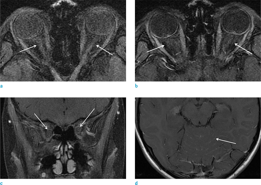
Fig. 3 Leukemic infiltration of bilateral optic nerves in a 10-year-old boy. He showed diplopia, which occurred 29 months after achieving a first complete remission of acute lymphoblastic leukemia. (a, b) Axial fat-suppressed, T1-weighted MR (a) and contrast-enhanced, fat-suppressed axial T1-weighted MR (b) images show subtle thickening with perineual enhancement of both optic nerves (arrows). (c) Contrast-enhanced, fat-suppressed coronal T1-weighted imaging also demonstrates mild neural thickening with perineural enhancement of both optic nerves (arrows). (d) Contrast-enhanced, axial T1-weighted image of the brain shows prominent leptomeningeal enhancement in the prepontine cistern and along the cerebellar folia (arrow).

Fig. 4 Right optic neuritis in an 11-year-old boy. He had a symptom of decreased visual acuity of right eye, which occurred 5 months after achieving a first complete remission in acute lymphoblastic leukemia. (a, b) Fat-suppressed axial T1-weighted MR (a) and contrast-enhanced, fat-suppressed axial T1-weighted MR (b) images demonstrate mild thickening and perineural enhancement of right optic nerve with mild intraconal fat infiltration (arrows).
Reference
-
1. Chen CY, Zimmerman RA, Faro S, Bilaniuk LT, Chou TY, Molloy PT. Childhood leukemia: central nervous system abnormalities during and after treatment. AJNR Am J Neuroradiol. 1996; 17:295–310.2. Brenner H, Kaatsch P, Burkhardt-Hammer T, Harms DO, Schrappe M, Michaelis J. Long-term survival of children with leukemia achieved by the end of the second millennium. Cancer. 2001; 92:1977–1983.3. Ginsberg LE, Leeds NE. Neuroradiology of leukemia. AJR Am J Roentgenol. 1995; 165:525–534.4. Camera A, Piccirillo G, Cennamo G, et al. Optic nerve involvement in acute lymphoblastic leukemia. Leuk Lymphoma. 1993; 11:153–155.5. Lin YC, Wang AG, Yen MY, Hsu WM. Leukaemic infiltration of the optic nerve as the initial manifestation of leukaemic relapse. Eye (Lond). 2004; 18:546–550.6. Madani A, Christophe C, Ferster A, Dan B. Peri-optic nerve infiltration during leukaemic relapse: MRI diagnosis. Pediatr Radiol. 2000; 30:30–32.7. Vazquez E, Lucaya J, Castellote A, et al. Neuroimaging in pediatric leukemia and lymphoma: differential diagnosis. Radiographics. 2002; 22:1411–1428.8. Arrigan M, Smyth L, Harmon M, Flynn C, Sheehy N. Imaging findings in recurrent extramedullary leukaemias. Cancer Imaging. 2013; 13:26–35.9. Porter RP, Kaste SC. Imaging findings of recurrent acute lymphoblastic leukemia in children and young adults, with emphasis on MRI. Pediatr Radiol. 2004; 34:400–408.10. de Fatima Soares M, Braga FT, da Rocha AJ, Lederman HM. Optic nerve infiltration by acute lymphoblastic leukemia: MRI contribution. Pediatr Radiol. 2005; 35:799–802.11. Rosenthal AR. Ocular manifestations of leukemia. A review. Ophthalmology. 1983; 90:899–905.12. Townsend JH, Dubovy SR, Pasol J, Lam BL. Transient optic perineuritis as the initial presentation of central nervous system involvement by pre-B cell lymphocytic leukemia. J Neuroophthalmol. 2013; 33:162–164.13. Puvanachandra N, Goddard K, Lyons CJ. Dramatic visual recovery after prompt radiotherapy and chemotherapy for leukaemic infiltration of the optic nerve in a child. Eye (Lond). 2010; 24:927–928.
