Hip Pelvis.
2016 Sep;28(3):173-177. 10.5371/hp.2016.28.3.173.
Pathologic Fracture of the Femur in Brown Tumor Induced in Parathyroid Carcinoma: A Case Report
- Affiliations
-
- 1Department of Urology and Endocrine Surgery, Haeundae Paik Hospital, Inje University College of Medicine, Busan, Korea.
- 2Department of Orthopaedic Surgery, Busan Paik Hospital, Inje University College of Medicine, Busan, Korea. docos@naver.com
- KMID: 2354550
- DOI: http://doi.org/10.5371/hp.2016.28.3.173
Abstract
- Brown tumor refers to a change of skeletones that develops as a complication of hyperparathyroidism. As osteoclast is activated to stimulate reabsorption and fibrosis of bone, it causes a cystic change of the bone. Parathyroid carcinoma is being reported as a tumor that induces primary hyperparathyroidism. It causes excessive secretion of the parathyroid hormone and increases the blood parathyroid hormone and calcium. Bone deformation due to brown tumor is known to be naturally recovered through the treatment for hyperparathyroidism. However, there is no clearly defined treatment for lesions that can induce pathological fractures developing in lower extremities. We experienced a case where brown tumor developed in the proximal femur of a 57-year-old female patient due to parathyroid carcinoma. In this case, spontaneous fracture occurred without any trauma, and it was cured by performing intramedullary nailing fixation and parathyroidectomy. We report the treatment results along with a literature review.
MeSH Terms
Figure
-
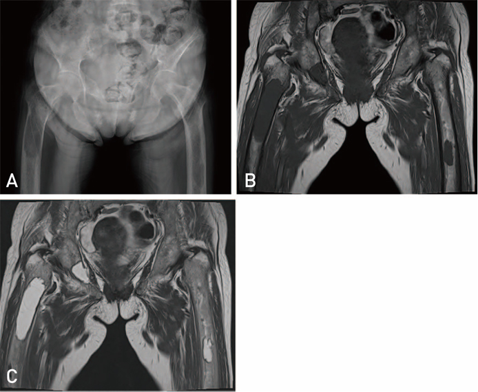
Fig. 1 X-ray findings at the time of hospital visit. (A) Osteolytic lesion in right proximal femur and left femur were observed. On pelvis magnetic resonance image, the signal of brown tumor was low in T1WI (B) and high in T2WI (C).
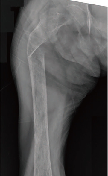
Fig. 2 Fracture of right proximal femur occurred without trauma.
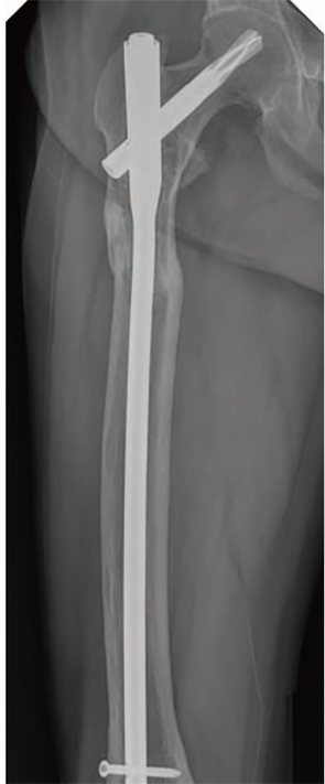
Fig. 3 The fracture on the right proximal femur was healed four months after the surgery.

Fig. 4 The final follow up X-ray shows healed fracture and improved multiple osteolytic lesion.
Reference
-
1. Ullah E, Ahmad M, Ali SA, Redhu N. Primary hyperparathyroidism having multiple Brown tumors mimicking malignancy. Indian J Endocrinol Metab. 2012; 16:1040–1042.
Article2. Bassler T, Wong ET, Brynes RK. Osteitis fibrosa cystica simulating metastatic tumor. An almost-forgotten relationship. Am J Clin Pathol. 1993; 100:697–700.
Article3. Radulescu D, Chis B, Donca V, Munteanu V. Brown tumors of the femur and pelvis secondary to a parathyroid carcinoma: report of one case. Rev Med Chil. 2014; 142:919–923.
Article4. Hsieh MC, Ko JY, Eng HL. Pathologic fracture of the distal femur in osteitis fibrosa cystica simulating metastatic disease. Arch Orthop Trauma Surg. 2004; 124:498–501.
Article5. Emin AH, Süogğlu Y, Demir D, Karatay MC. Normocalcemic hyperparathyroidism presented with mandibular brown tumor: report of a case. Auris Nasus Larynx. 2004; 31:299–304.
Article6. Hoshi M, Takami M, Kajikawa M, et al. A case of multiple skeletal lesions of brown tumors, mimicking carcinoma metastases. Arch Orthop Trauma Surg. 2008; 128:149–154.
Article7. Jawad MU, Scully SP. In brief: classifications in brief: Mirels' classification: metastatic disease in long bones and impending pathologic fracture. Clin Orthop Relat Res. 2010; 468:2825–2827.8. Mirels H. Metastatic disease in long bones. A proposed scoring system for diagnosing impending pathologic fractures. Clin Orthop Relat Res. 1989; (249):256–264.
Article
- Full Text Links
-
- Actions
-
Cited
- CITED
-
- Close
- Share
-
- Similar articles
-
- Brown Tumor of The Spine with Compression Fracture: A Case Report
- A Case of Parathyroid Carcinoma Presenting as Brown Tumors
- A case of mediastinal parathyroid adenoma presenting as fracture of brown tumor
- Intermittent Parathyroid Hormone Treatment for Stimulation of Callus Formation on Distal Femoral Fracture in Elderly Patients: Case Report
- Paget's Disease of Bone with Pathologic Fracture of the Femur: a Case Report
