J Korean Soc Radiol.
2016 Oct;75(4):322-326. 10.3348/jksr.2016.75.4.322.
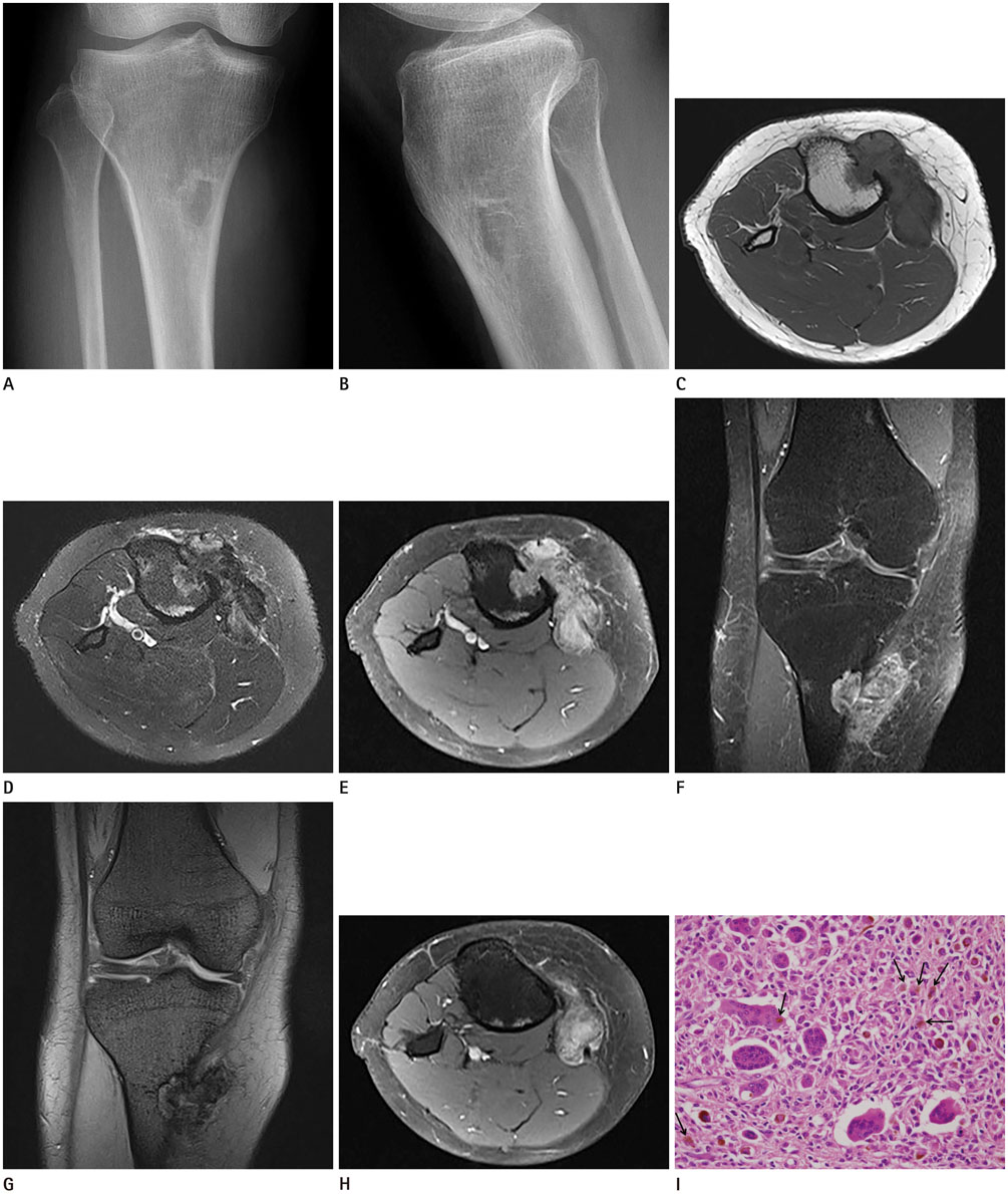
Tenosynovial Giant Cell Tumor of the Pes Anserine Bursa with Bone Marrow Extension into the Tibia: A Case Report
- Affiliations
-
- 1Department of Radiology, St. Vincent's Hospital, College of Medicine, The Catholic University of Korea, Suwon, Korea. jeeykim@catholic.ac.kr
- 2Department of Orthopedic Surgery, St. Vincent's Hospital, College of Medicine, The Catholic University of Korea, Suwon, Korea.
- 3Department of Hospital Pathology, St. Vincent's Hospital, College of Medicine, The Catholic University of Korea, Suwon, Korea.
- KMID: 2353685
- DOI: http://doi.org/10.3348/jksr.2016.75.4.322
Abstract
- Extra-articular diffuse-type tenosynovial giant cell tumor (TSGCT), especially arising from the pes anserine bursa, is a rare lesion. Only one case of extra-articular diffuse-type TSGCT of the pes anserine bursa that showed bone erosion as an imaging finding has been reported. Here, we report our experience of an exceptional case of extra-articular diffuse-type TSGCT of the pes anserine bursa with intramedullary extension into the tibia on magnetic resonance imaging, which resembled a benign soft tissue mass on radiography, along with a review of the literature.
MeSH Terms
Figure
Reference
-
1. Sun C, Sheng W, Yu H, Han J. Giant cell tumor of the tendon sheath: a rare case in the left knee of a 15-year-old boy. Oncol Lett. 2012; 3:718–720.2. Jaffe HL, Lichtenstein L, Sutro CJ. Pigmented villonodular synovitis, bursitis and tenosynovitis: a discussion of the synovial and bursal equivalents of the tenosynovial lesion commonly denoted as xanthoma, xanthogranuloma, giant cell tumor or myeloplaxoma of the tendon sheath, with some consideration of this tendon sheath lesion itself. Arch Pathol. 1941; 31:731–765.3. Lucas DR. Tenosynovial giant cell tumor: case report and review. Arch Pathol Lab Med. 2012; 136:901–906.4. Zhao H, Maheshwari AV, Kumar D, Malawer MM. Giant cell tumor of the pes anserine bursa (extra-articular pigmented villonodular bursitis): a case report and review of the literature. Case Rep Med. 2011; 2011:491470.5. Present DA, Bertoni F, Enneking WF. Case report 348: pigmented villonodular synovitis arising from bursa of the pes anserinus muscle, with secondary involvement of the tibia. Skeletal Radiol. 1986; 15:236–240.6. Maheshwari AV, Muro-Cacho CA, Pitcher JD Jr. Pigmented villonodular bursitis/diffuse giant cell tumor of the pes anserine bursa: a report of two cases and review of literature. Knee. 2007; 14:402–407.7. Richman DM, Bresler SC, Rosenthal MH, Howard SA. Malignant tenosynovial giant cell tumor of the leg: a radiologic-pathologic correlation and review of the literature. J Clin Imaging Sci. 2015; 5:13.8. Middleton WD, Patel V, Teefey SA, Boyer MI. Giant cell tumors of the tendon sheath: analysis of sonographic findings. AJR Am J Roentgenol. 2004; 183:337–339.9. Sheldon PJ, Forrester DM, Learch TJ. Imaging of intraarticular masses. Radiographics. 2005; 25:105–119.10. Zeiss J, Coombs RJ, Booth RL Jr, Saddemi SR. Chronic bursitis presenting as a mass in the pes anserine bursa: MR diagnosis. J Comput Assist Tomogr. 1993; 17:137–140.
- Full Text Links
- Actions
-
Cited
- CITED
-
- Close
- Share
- Similar articles
-
- Sonoanatomic Variation of Pes Anserine Bursa
- Pes anserinus and anserine bursa: anatomical study
- Tenosynovial Giant Cell Tumor Showing Severe Bone Erosion in the Finger: Case Report and Review of the Imaging Findings and Their Significance
- Tenosynovial Giant-cell Tumor of the Lumbar Spine
- Tenosynovial Giant Cell Tumor of the Temporomandibular Joint


