J Pathol Transl Med.
2016 Sep;50(5):394-396. 10.4132/jptm.2016.03.11.
Nodular Fasciitis of External Auditory Canal
- Affiliations
-
- 1Department of Pathology, Dongkang Medical Center, Ulsan, Korea. jvcjh@hanmail.net
- 2Department of Otolaryngology, Dongkang Medical Center, Ulsan, Korea.
- KMID: 2353603
- DOI: http://doi.org/10.4132/jptm.2016.03.11
Abstract
- Nodular fasciitis is a pseudosarcomatous reactive process composed of fibroblasts and myofibroblasts, and it is most common in the upper extremities. Nodular fasciitis of the external auditory canal is rare. To the best of our knowledge, less than 20 cases have been reported to date. We present a case of nodular fasciitis arising in the cartilaginous part of the external auditory canal. A 19-year-old man complained of an auricular mass with pruritus. Computed tomography showed a 1.7 cm sized soft tissue mass in the right external auditory canal, and total excision was performed. Histologic examination revealed spindle or stellate cells proliferation in a fascicular and storiform pattern. Lymphoid cells and erythrocytes were intermixed with tumor cells. The stroma was myxoid to hyalinized with a few microcysts. The tumor cells were immunoreactive for smooth muscle actin, but not for desmin, caldesmon, CD34, S-100, anaplastic lymphoma kinase, and cytokeratin. The patient has been doing well during the 1 year follow-up period.
Keyword
MeSH Terms
Figure
-
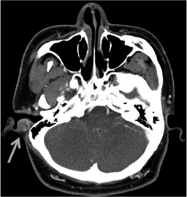
Fig. 1. Radiologic finding. An ovoid mass with soft tissue density (arrow) is noted in the right external auditory canal on computed tomography.

Fig. 2. Pathologic findings. (A) Spindle cell proliferation shows a vaguely storiform to fascicular pattern. (B) The bland spindle and stellate cells are set in loose myxoid (upper portion) to hyalinized matrix (lower portion). Extravasated red blood cells and scattered lymphoid cells are identified. (C) The tumor cells are positive for smooth muscle actin.
Reference
-
1. Konwaler BE, Keasbey L, Kaplan L. Subcutaneous pseudosarcomatous fibromatosis (fasciitis). Am J Clin Pathol. 1955; 25:241–52.
Article2. Weiss SW, Goldblum JR. Enzinger and Weiss’s soft tissue tumors. 5th ed. St. Louis: Mosby;2008.3. Thompson LD, Fanburg-Smith JC, Wenig BM. Nodular fasciitis of the external ear region: a clinicopathologic study of 50 cases. Ann Diagn Pathol. 2001; 5:191–8.
Article4. Abdel-Aziz M, Khattab H, El-bosraty H, El-hoshy H, Hesham A, Al-taweel HW. Nodular fasciitis of the external auditory canal in six Egyptian children. Int J Pediatr Otorhinolaryngol. 2008; 72:643–6.
Article5. Peng WX, Kudo M, Yamamoto T, et al. Nodular fasciitis in the parotid gland: a case report and review of the literature. Diagn Cytopathol. 2013; 41:829–33.
Article6. Jung KH, Kim YW, So YK, Choi SI, Baek MJ. Inflammatory myofibroblastic tumor involving ear lobule. Auris Nasus Larynx. 2012; 39:631–3.
Article7. Bhattacharya B, Dilworth HP, Iacobuzio-Donahue C, et al. Nuclear beta-catenin expression distinguishes deep fibromatosis from other benign and malignant fibroblastic and myofibroblastic lesions. Am J Surg Pathol. 2005; 29:653–9.8. Morrissey G, Robinson AC, Stirling R. Cellular benign fibrous histiocytoma of the external auditory meatus. J Laryngol Otol. 1996; 110:98–100.
Article9. Rezk S, Yousef M, Zamansky M, Khan A. Solitary fibrous tumor of the auditory canal. Arch Pathol Lab Med. 2004; 128:e169–71.
Article10. Kim JR, Chi JG. Nodular fasciitis (13 cases analysis). Korean J Pathol. 1998; 22:190–4.11. Meng GZ, Zhang HY, Zhang Z, Wei B, Bu H. Myofibroblastic sarcoma vs nodular fasciitis: a comparative study of chromosomal imbalances. Am J Clin Pathol. 2009; 131:701–9.12. Erickson-Johnson MR, Chou MM, Evers BR, et al. Nodular fasciitis: a novel model of transient neoplasia induced by MYH9-USP6 gene fusion. Lab Invest. 2011; 91:1427–33.13. Amary MF, Ye H, Berisha F, Tirabosco R, Presneau N, Flanagan AM. Detection of USP6 gene rearrangement in nodular fasciitis: an important diagnostic tool. Virchows Arch. 2013; 463:97–8.14. Oliveira AM, Chou MM. USP6-induced neoplasms: the biologic spectrum of aneurysmal bone cyst and nodular fasciitis. Hum Pathol. 2014; 45:1–11.
Article
- Full Text Links
-
- Actions
-
Cited
- CITED
-
- Close
- Share
-
- Similar articles
-
- A Case of Osteoma with Cholesteatoma in the External Auditory Canal
- Four Cases of the External Auditory Canal Cancer
- A Case of Langerhans Cell Histiocytosis involving the External Auditory Canal
- Compound Nevus Occurring Near External Auditory Canal: Successful Treatment by CO2 Laser Abrasion
- A Case of Nodular Pseudosarcomatous Fasciitis
