Mammary-Type Myofibroblastoma: A Report of Two Cases
- Affiliations
-
- 1Department of Pathology, Asan Medical Center, University of Ulsan College of Medicine, Seoul, Korea. Songjs@amc.seoul.kr
- 2Department of Radiology, Kyung Hee University Hospital at Gangdong, Kyung Hee University School of Medicine, Seoul, Korea.
- 3Department of Surgery, Asan Medical Center, University of Ulsan College of Medicine, Seoul, Korea.
- KMID: 2353601
- DOI: http://doi.org/10.4132/jptm.2016.03.26
Abstract
- Mammary-type myofibroblastoma (MFB) is a rare, benign spindle cell neoplasm occurring along the milkline, with extension from the mid-axilla to the medial groin. It is histologically and immunohistochemically identical to MFB of the breast and is part of a spectrum of lesions that includes spindle cell lipoma and cellular angiofibroma. Recently, we experienced two cases of mammary-type MFB involving male patients aged 30 and 58 years, respectively. The tumors were located in the right scrotal sac and in the right axilla. Wide excisions were performed. Microscopically, the masses were composed of haphazardly arranged, variably sized fascicles of bland spindle cells and were admixed with mature fat tissue. The spindle cells in both cases showed immunopositivity for desmin and CD34 and negativity for smooth muscle actin. Loss of retinoblastoma (RB)/13q14 loci is a characteristic genetic alteration of mammary-type MFB, and we identified loss of RB protein expression by immunohistochemical staining. We emphasize the importance of awareness of this rare neoplasm when a spindle cell neoplasm is accompanied by desmin immunopositivity. The second patient was alive without recurrence for 20 months, and the first patient had not been followed.
Keyword
MeSH Terms
Figure
-
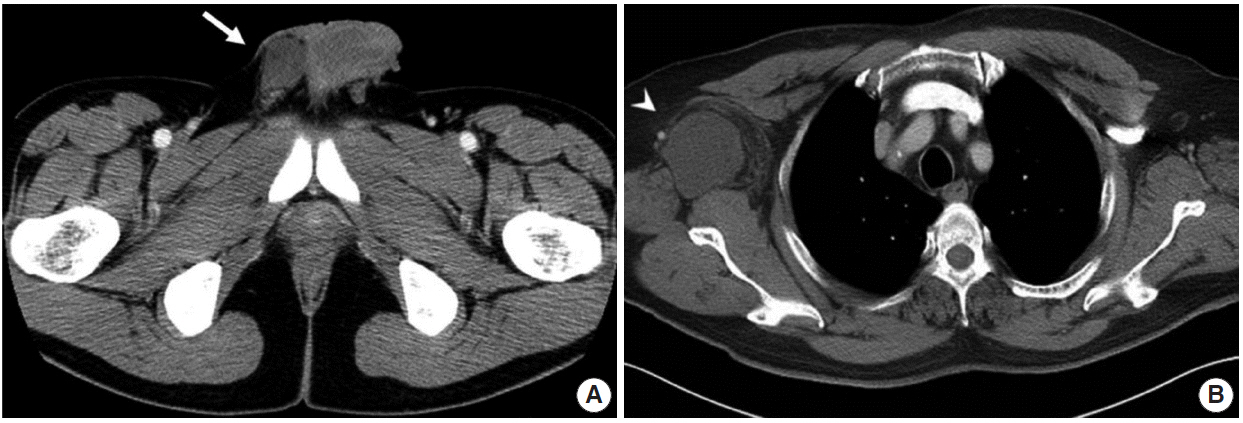
Fig. 1. Radiologic findings for the two study cases. (A) In case 1, a pelvis computed tomography (CT) shows 5.0 cm mass in the right scrotal sac separated from the adjacent testis and spermatic cord (arrow). (B) In case 2, a chest CT shows a 5.5 cm lobulated mass in the right axilla (arrowhead).

Fig. 2. Gross and microscopic findings of case 1. (A) The cut surface is yellowish tan, rubbery, and trabeculated, without necrosis or hemorrhage. (B) The mass is composed of haphazardly arranged and variably sized fascicles of bland spindle cells. Tumor cells have tapered nuclei and fine chromatin. Numerous mast cells are scattered (arrows). No mitosis is observed. The tumor cells show immunopositivity for desmin (C) and CD34 (D).
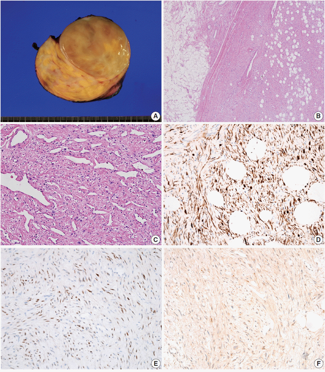
Fig. 3. Gross and microscopic findings of case 2. (A) The cut surface is yellowish tan, smooth, and glistening, and a focal myxoid change is observed. (B) The mass is well demarcated with mature adipocytes. (C) Staghorn-like blood vessels are present, with focal hyalinization. Tumor cells shows immunopositivity for desmin (D) and estrogen receptor (E). (F) Retinoblastoma immunostaining shows negativity.
Reference
-
1. Hameed M, Clarke K, Amer HZ, Mahmet K, Aisner S. Cellular angiofibroma is genetically similar to spindle cell lipoma: a case report. Cancer Genet Cytogenet. 2007; 177:131–4.
Article2. McMenamin ME, Fletcher CD. Mammary-type myofibroblastoma of soft tissue: a tumor closely related to spindle cell lipoma. Am J Surg Pathol. 2001; 25:1022–9.3. Mukonoweshuro P, McCormick F, Rachapalli V, Natale S, Smith ME. Paratesticular mammary-type myofibroblastoma. Histopathology. 2007; 50:396–7.
Article4. Arsenovic N, Abdulla KE, Shamim KS. Mammary-type myofibroblastoma of soft tissue. Indian J Pathol Microbiol. 2011; 54:391–3.
Article5. Millo NZ, Yee EU, Mortele KJ. Mammary-type myofibroblastoma of the liver: multi-modality imaging features with histopathologic correlation. Abdom Imaging. 2014; 39:482–7.
Article6. Howitt BE, Fletcher CD. Mammary-type myofibroblastoma: clinicopathologic characterization in a series of 143 cases. Am J Surg Pathol. 2016; 40:361–7.7. Fletcher CD, Bridge JA, Hogendoorn P, Mertens F. WHO classification of tumours of soft tissue and bone. Lyon: IARC Press;2013. p. 61–2.8. Hox V, Vander Poorten V, Delaere PR, Hermans R, Debiec-Rychter M, Sciot R. Extramammary myofibroblastoma in the head and neck region. Head Neck. 2009; 31:1240–4.
Article9. Maggiani F, Debiec-Rychter M, Verbeeck G, Sciot R. Extramammary myofibroblastoma is genetically related to spindle cell lipoma. Virchows Arch. 2006; 449:244–7.
Article10. Flucke U, van Krieken JH, Mentzel T. Cellular angiofibroma: analysis of 25 cases emphasizing its relationship to spindle cell lipoma and mammary-type myofibroblastoma. Mod Pathol. 2011; 24:82–9.
Article11. Fritchie KJ, Carver P, Sun Y, et al. Solitary fibrous tumor: is there a molecular relationship with cellular angiofibroma, spindle cell lipoma, and mammary-type myofibroblastoma? Am J Clin Pathol. 2012; 137:963–70.12. Magro G, Righi A, Casorzo L, et al. Mammary and vaginal myofibroblastomas are genetically related lesions: fluorescence in situ hybridization analysis shows deletion of 13q14 region. Hum Pathol. 2012; 43:1887–93.
Article13. Magro G. Chromosome 13q14 deletion in a mammary-type myofibroblastoma of the big toe: reply. Hum Pathol. 2015; 46:344–5.14. Gengler C, Guillou L. Solitary fibrous tumour and haemangiopericytoma: evolution of a concept. Histopathology. 2006; 48:63–74.
Article15. Meguerditchian AN, Malik DA, Hicks DG, Kulkarni S. Solitary fibrous tumor of the breast and mammary myofibroblastoma: the same lesion? Breast J. 2008; 14:287–92.
Article
- Full Text Links
-
- Actions
-
Cited
- CITED
-
- Close
- Share
-
- Similar articles
-
- Epithelioid Myofibroblastoma of Mammary-type in Chest Wall: A Case Report
- A Case of Myofibroblastoma in the Submandibular Region
- A Case of Mammary-Type Myofibroblastoma of the Tongue
- A Mammary Type Myofibroblastoma in the Pelvic Cavity: A Case Report
- Mammary-Type Myofibroblastoma of the Buttock: A Case Report and Review of the Literature
