J Korean Soc Radiol.
2017 Feb;76(2):142-147. 10.3348/jksr.2017.76.2.142.
Mammary-Type Myofibroblastoma of the Buttock: A Case Report and Review of the Literature
- Affiliations
-
- 1Department of Radiology, Kyungpook National University Hospital, College of Medicine, Kyungpook National University, Daegu, Korea. yijh72@gmail.com
- KMID: 2367862
- DOI: http://doi.org/10.3348/jksr.2017.76.2.142
Abstract
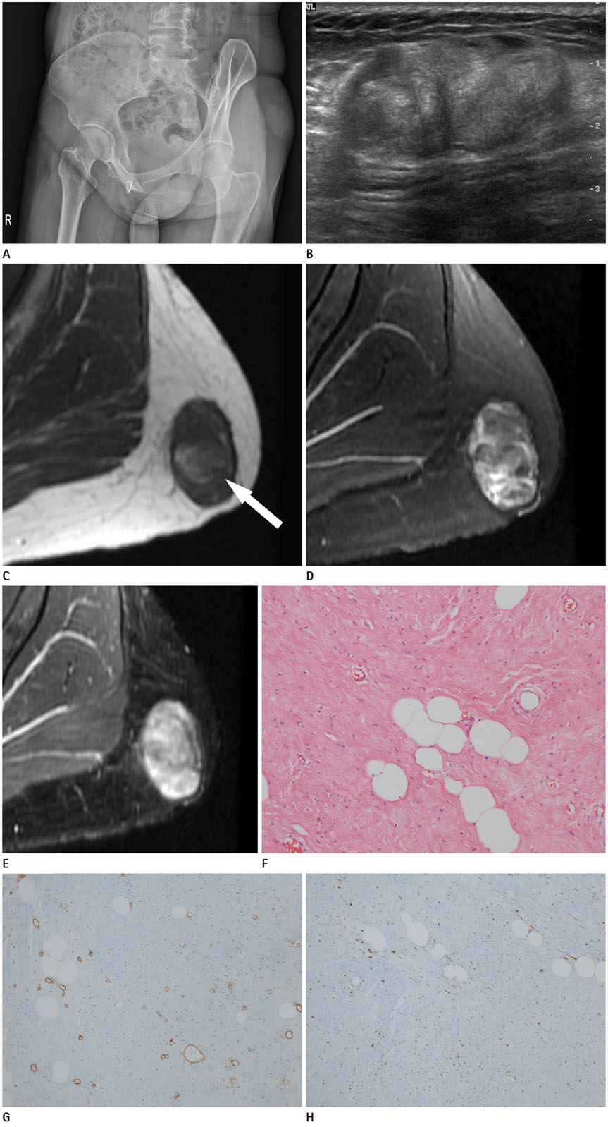
- Mammary-type myofibroblastoma is a very rare, benign mesenchymal tumor consisting of spindle-shaped cells along with thick hyalinized collagen bundles and an intralesional fat component; its histopathological features are identical to those of myofibroblastomas of the breast. It usually occurs along the embryonic milk-line; however, unusual cases occurring outside of the embryonic milk-line have also been reported. Although this tumor always shows clinically benign behavior, its variable histological composition can easily be confused with many other fibrous and lipomatous neoplasms. Unfortunately, its radiological findings are extremely rarely described in the literature. Here, we present a rare case of mammary-type myofibroblastoma in a 38-year-old woman who presented with a well-circumscribed solitary mass in the buttock, and discuss various radiologic imaging findings, such as plain radiography, ultrasonography, and magnetic resonance imaging results.
MeSH Terms
Figure
Reference
-
1. Wargotz ES, Weiss SW, Norris HJ. Myofibroblastoma of the breast. Sixteen cases of a distinctive benign mesenchymal tumor. Am J Surg Pathol. 1987; 11:493–502.2. McMenamin ME, Fletcher CD. Mammary-type myofibroblastoma of soft tissue: a tumor closely related to spindle cell lipoma. Am J Surg Pathol. 2001; 25:1022–1029.3. Bhullar JS, Varshney N, Dubay L. Intranodal palisaded myofibroblastoma: a review of the literature. Int J Surg Pathol. 2013; 21:337–341.4. Abdul-Ghafar J, Ud Din N, Ahmad Z, Billings SD. Mammary-type myofibroblastoma of the right thigh: a case report and review of the literature. J Med Case Rep. 2015; 9:126.5. Scotti C, Camnasio F, Rizzo N, Fontana F, De Cobelli F, Peretti GM, et al. Mammary-type myofibroblastoma of popliteal fossa. Skeletal Radiol. 2008; 37:549–553.6. Millo NZ, Yee EU, Mortele KJ. Mammary-type myofibroblastoma of the liver: multi-modality imaging features with histopathologic correlation. Abdom Imaging. 2014; 39:482–487.7. Agale SV, Warpe BM, Kumari G, Valand AG. Myofibroblastoma of axillary soft tissue in a child. J Cancer Tumor Int. 2015; 2:150–154.8. Bigotti G, Coli A, Mottolese M, Di Filippo F. Selective location of palisaded myofibroblastoma with amianthoid fibres. J Clin Pathol. 1991; 44:761–764.9. Magro G. Mammary myofibroblastoma: a tumor with a wide morphologic spectrum. Arch Pathol Lab Med. 2008; 132:1813–1820.10. Yoo EY, Shin JH, Ko EY, Han BK, Oh YL. Myofibroblastoma of the female breast: mammographic, sonographic, and magnetic resonance imaging findings. J Ultrasound Med. 2010; 29:1833–1836.


