Finite Element Analysis for Comparison of Spinous Process Osteotomies Technique with Conventional Laminectomy as Lumbar Decompression Procedure
- Affiliations
-
- 1Spine Center and Department of Orthopedic Surgery, Seoul National University College of Medicine, Seoul National University Bundang Hospital, Seongnam, Korea. highcervical@gmail.com
- 2Department of Mechanical Engineering, Yonsei University, Seoul, Korea.
- 3Department of Orthopedic Surgery, Yonsei University College of Medicine, Seoul, Korea.
- 4Department of Orthopedic Surgery, Seoul National University College of Medicine, Seoul National University Hospital, Seoul, Korea.
- KMID: 2352800
- DOI: http://doi.org/10.3349/ymj.2015.56.1.146
Abstract
- PURPOSE
The purpose of this study was to evaluate and compare the biomechanical behavior of the lumbar spine after posterior decompression with the spinous process osteotomy (SPiO) technique or the conventional laminectomy (CL) technique using a finite element (FE) model.
MATERIALS AND METHODS
Three validated lumbar FE models (L2-5) which represented intact spine and two decompression models using SPiO and CL techniques at the L3-4 segment were developed. In each model, the ranges of motion, the maximal von Mises stress of the annulus fibrosus, and the intradiscal pressures at the index segment (L3-4) and adjacent segments (L2-3 and L4-5) under 7.5 Nm moments were analyzed. Facet contact forces were also compared among three models under the extension and torsion moments.
RESULTS
Compared to the intact model, the CL and SPiO models had increased range of motion and annulus stress at both the index segment (L3-4) and the adjacent segments under flexion and torsion. However, the SPiO model demonstrated a reduced range of motion and annulus stress than the CL model. Both CL and SPiO models had an increase of facet contact force at the L3-4 segment under the torsion moment compared to that of the intact model. Under the extension moment, however, three models demonstrated a similar facet contact force even at the L3-4 model.
CONCLUSION
Both decompression methods lead to postoperative segmental instability compared to the intact model. However, SPiO technique leads to better segmental stability compared to the CL technique.
Keyword
MeSH Terms
-
Biomechanical Phenomena
Decompression, Surgical/*methods
*Finite Element Analysis
Humans
Intervertebral Disc/physiopathology/surgery
Laminectomy/*methods
Lumbar Vertebrae/pathology/physiopathology/*surgery
Male
Middle Aged
Models, Anatomic
Osteotomy/*methods
Range of Motion, Articular
Stress, Mechanical
Zygapophyseal Joint/pathology/physiopathology/surgery
Figure
-

Fig. 1 Finite element models. (A) Conventional laminectomy (CL) model; arrow indicates the sacrifice of posterior ligament complex. (B) Spinous process osteotomy (SPiO) model; arrow indicates the preservation of posterior ligament complex.
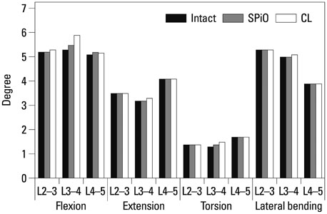
Fig. 2 Comparison between the three models of the range of motion at each segment for the four moments. CL, conventional laminectomy; SPiO, spinous process osteotomy.

Fig. 3 Comparison of the maximal von Mises stress of the annulus fibrosus and the intradiscal pressure of the nucleus pulposus among three models. (A) Comparison of the maximal von Mises stress of the annulus fibrosus (MPa). (B) Comparison of intradiscal pressure of the nucleus pulposus.
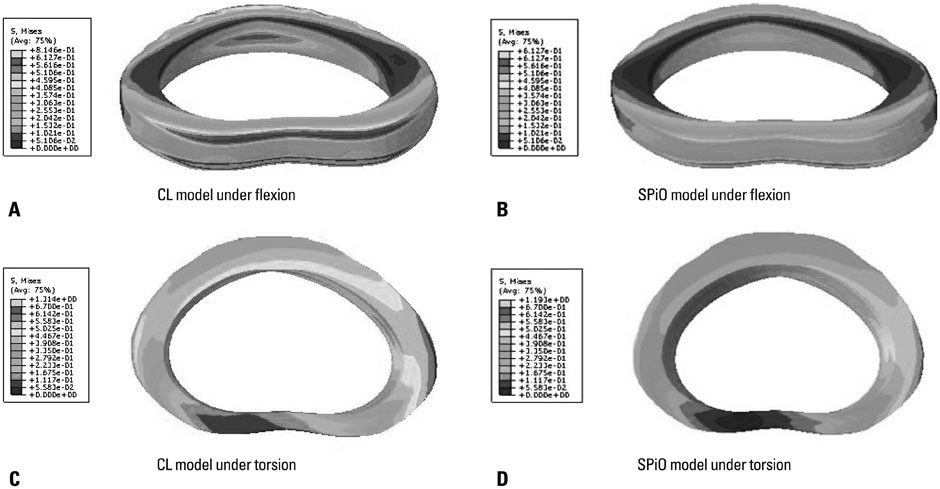
Fig. 4 Distribution of stress of the annulus fibrosus in the CL and SPiO models under flexion and torsion moment. (A) CL model under flexion. (B) SPiO model under flexion. (C) CL model under torsion. (D) SPiO model under torsion. CL, conventional laminectomy; SPiO, spinous process osteotomy.

Fig. 5 Comparison of the facet contact force among 3 models. CL, conventional laminectomy; SPiO, spinous process osteotomy.
Reference
-
1. Verbiest H. A radicular syndrome from developmental narrowing of the lumbar vertebral canal. J Bone Joint Surg Br. 1954; 36-B:230–237.2. Hilibrand AS, Rand N. Degenerative lumbar stenosis: diagnosis and management. J Am Acad Orthop Surg. 1999; 7:239–249.
Article3. Sengupta DK, Herkowitz HN. Lumbar spinal stenosis. Treatment strategies and indications for surgery. Orthop Clin North Am. 2003; 34:281–295.4. Atlas SJ, Keller RB, Wu YA, Deyo RA, Singer DE. Long-term outcomes of surgical and nonsurgical management of lumbar spinal stenosis: 8 to 10 year results from the maine lumbar spine study. Spine (Phila Pa 1976). 2005; 30:936–943.
Article5. Bresnahan L, Ogden AT, Natarajan RN, Fessler RG. A biomechanical evaluation of graded posterior element removal for treatment of lumbar stenosis: comparison of a minimally invasive approach with two standard laminectomy techniques. Spine (Phila Pa 1976). 2009; 34:17–23.
Article6. Ikuta K, Tono O, Tanaka T, Arima J, Nakano S, Sasaki K, et al. Surgical complications of microendoscopic procedures for lumbar spinal stenosis. Minim Invasive Neurosurg. 2007; 50:145–149.
Article7. Yuzawa Y. The interspinous ligament should be removed for the decompression surgery with the case of lumbar spinal canal stenosis. Arch Orthop Trauma Surg. 2011; 131:753–758.
Article8. Weiner BK, Fraser RD, Peterson M. Spinous process osteotomies to facilitate lumbar decompressive surgery. Spine (Phila Pa 1976). 1999; 24:62–66.
Article9. El-Abed K, Barakat M, Ainscow D. Multilevel lumbar spinal stenosis decompression: midterm outcome using a modified hinge osteotomy technique. J Spinal Disord Tech. 2011; 24:376–380.10. Shirazi-Adl SA, Shrivastava SC, Ahmed AM. Stress analysis of the lumbar disc-body unit in compression. A three-dimensional nonlinear finite element study. Spine (Phila Pa 1976). 1984; 9:120–134.
Article11. Pintar FA, Yoganandan N, Myers T, Elhagediab A, Sances A Jr. Biomechanical properties of human lumbar spine ligaments. J Biomech. 1992; 25:1351–1356.
Article12. Goel VK, Kim YE, Lim TH, Weinstein JN. An analytical investigation of the mechanics of spinal instrumentation. Spine (Phila Pa 1976). 1988; 13:1003–1011.
Article13. Wu HC, Yao RF. Mechanical behavior of the human annulus fibrosus. J Biomech. 1976; 9:1–7.
Article14. Shirazi-Adl A, Ahmed AM, Shrivastava SC. Mechanical response of a lumbar motion segment in axial torque alone and combined with compression. Spine (Phila Pa 1976). 1986; 11:914–927.
Article15. Polikeit A, Ferguson SJ, Nolte LP, Orr TE. Factors influencing stresses in the lumbar spine after the insertion of intervertebral cages: finite element analysis. Eur Spine J. 2003; 12:413–420.
Article16. Renner SM, Natarajan RN, Patwardhan AG, Havey RM, Voronov LI, Guo BY, et al. Novel model to analyze the effect of a large compressive follower pre-load on range of motions in a lumbar spine. J Biomech. 2007; 40:1326–1332.
Article17. Schilling C, Krüger S, Grupp TM, Duda GN, Blömer W, Rohlmann A. The effect of design parameters of dynamic pedicle screw systems on kinematics and load bearing: an in vitro study. Eur Spine J. 2011; 20:297–307.
Article18. Wilson DC, Niosi CA, Zhu QA, Oxland TR, Wilson DR. Accuracy and repeatability of a new method for measuring facet loads in the lumbar spine. J Biomech. 2006; 39:348–353.
Article19. Kim HJ, Chun HJ, Lee HM, Kang KT, Lee CK, Chang BS, et al. The biomechanical influence of the facet joint orientation and the facet tropism in the lumbar spine. Spine J. 2013; 13:1301–1308.
Article20. Patwardhan AG, Havey RM, Meade KP, Lee B, Dunlap B. A follower load increases the load-carrying capacity of the lumbar spine in compression. Spine (Phila Pa 1976). 1999; 24:1003–1009.
Article21. Goel VK, Fromknecht SJ, Nishiyama K, Weinstein J, Liu YK. The role of lumbar spinal elements in flexion. Spine (Phila Pa 1976). 1985; 10:516–523.
Article22. Hindle RJ, Pearcy MJ, Cross A. Mechanical function of the human lumbar interspinous and supraspinous ligaments. J Biomed Eng. 1990; 12:340–344.
Article23. Gillespie KA, Dickey JP. Biomechanical role of lumbar spine ligaments in flexion and extension: determination using a parallel linkage robot and a porcine model. Spine (Phila Pa 1976). 2004; 29:1208–1216.
Article24. Natarajan RN, Andersson GB, Patwardhan AG, Andriacchi TP. Study on effect of graded facetectomy on change in lumbar motion segment torsional flexibility using three-dimensional continuum contact representation for facet joints. J Biomech Eng. 1999; 121:215–221.
Article25. Lee KK, Teo EC, Qiu TX, Yang K. Effect of facetectomy on lumbar spinal stability under sagittal plane loadings. Spine (Phila Pa 1976). 2004; 29:1624–1631.
Article26. Adams MA, Hutton WC. The effect of posture on the role of the apophysial joints in resisting intervertebral compressive forces. J Bone Joint Surg Br. 1980; 62:358–362.
Article27. Bydon A, Xu R, Parker SL, McGirt MJ, Bydon M, Gokaslan ZL, et al. Recurrent back and leg pain and cyst reformation after surgical resection of spinal synovial cysts: systematic review of reported postoperative outcomes. Spine J. 2010; 10:820–826.
Article28. Xu R, McGirt MJ, Parker SL, Bydon M, Olivi A, Wolinsky JP, et al. Factors associated with recurrent back pain and cyst recurrence after surgical resection of one hundred ninety-five spinal synovial cysts: analysis of one hundred sixty-seven consecutive cases. Spine (Phila Pa 1976). 2010; 35:1044–1053.
Article29. Kirkaldy-Willis WH, Farfan HF. Instability of the lumbar spine. Clin Orthop Relat Res. 1982; 110–123.
Article30. Galbusera F, Schmidt H, Neidlinger-Wilke C, Gottschalk A, Wilke HJ. The mechanical response of the lumbar spine to different combinations of disc degenerative changes investigated using randomized poroelastic finite element models. Eur Spine J. 2011; 20:563–571.
Article
- Full Text Links
-
- Actions
-
Cited
- CITED
-
- Close
- Share
-
- Similar articles
-
- Erratum to "Finite Element Analysis for Comparison of Spinous Process Osteotomies Technique with Conventional Laminectomy as Lumbar Decompression Procedure" by Kim HJ, et al. (Yonsei Med J 2015;56:146-53.)
- Comparison of Spinous Process-Splitting Laminectomy versus Conventional Laminectomy for Lumbar Spinal Stenosis
- Restoration of the Spinous Process Following Muscle-Preserving Posterior Lumbar Decompression via Sagittal Splitting of the Spinous Process
- Microsurgical Decompression of Lumbar Stenosis: Technical Innovations and Early Experience of 20 Cases
- Comparison of the Results of the Decompression Methods for Degenerative Lumbar Spinal Stenosis: Comparison of Posterior Element Saving Procedures
