J Adv Prosthodont.
2016 Aug;8(4):275-284. 10.4047/jap.2016.8.4.275.
Microtensile bond strength and micromorphologic analysis of surface-treated resin nanoceramics
- Affiliations
-
- 1Department of Prosthodontics, College of Dentistry, Dankook University, Cheonan, Republic of Korea. yu0324@hanmail.net
- KMID: 2349335
- DOI: http://doi.org/10.4047/jap.2016.8.4.275
Abstract
- PURPOSE
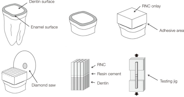
The aim of this study was to evaluate the influence of different surface treatment methods on the microtensile bond strength of resin cement to resin nanoceramic (RNC).
MATERIALS AND METHODS
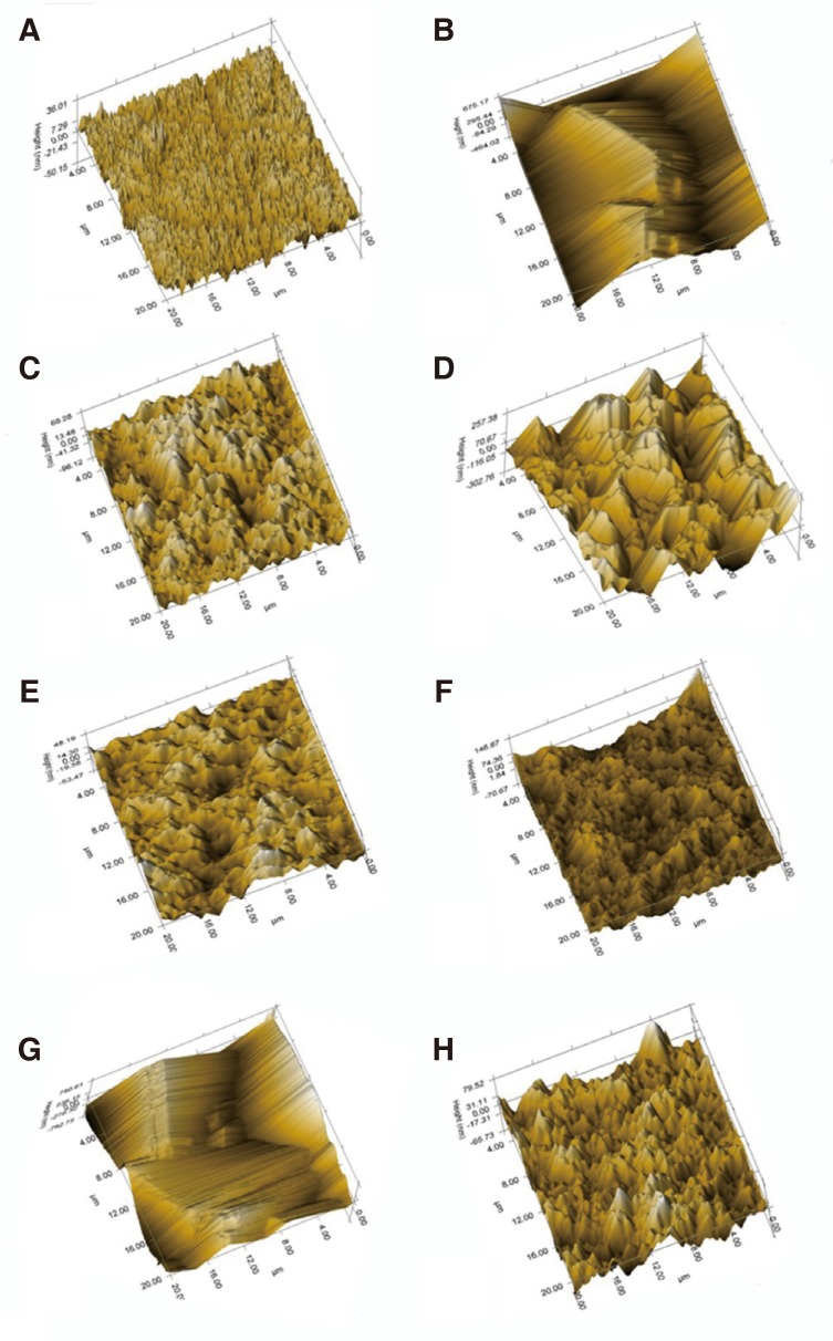
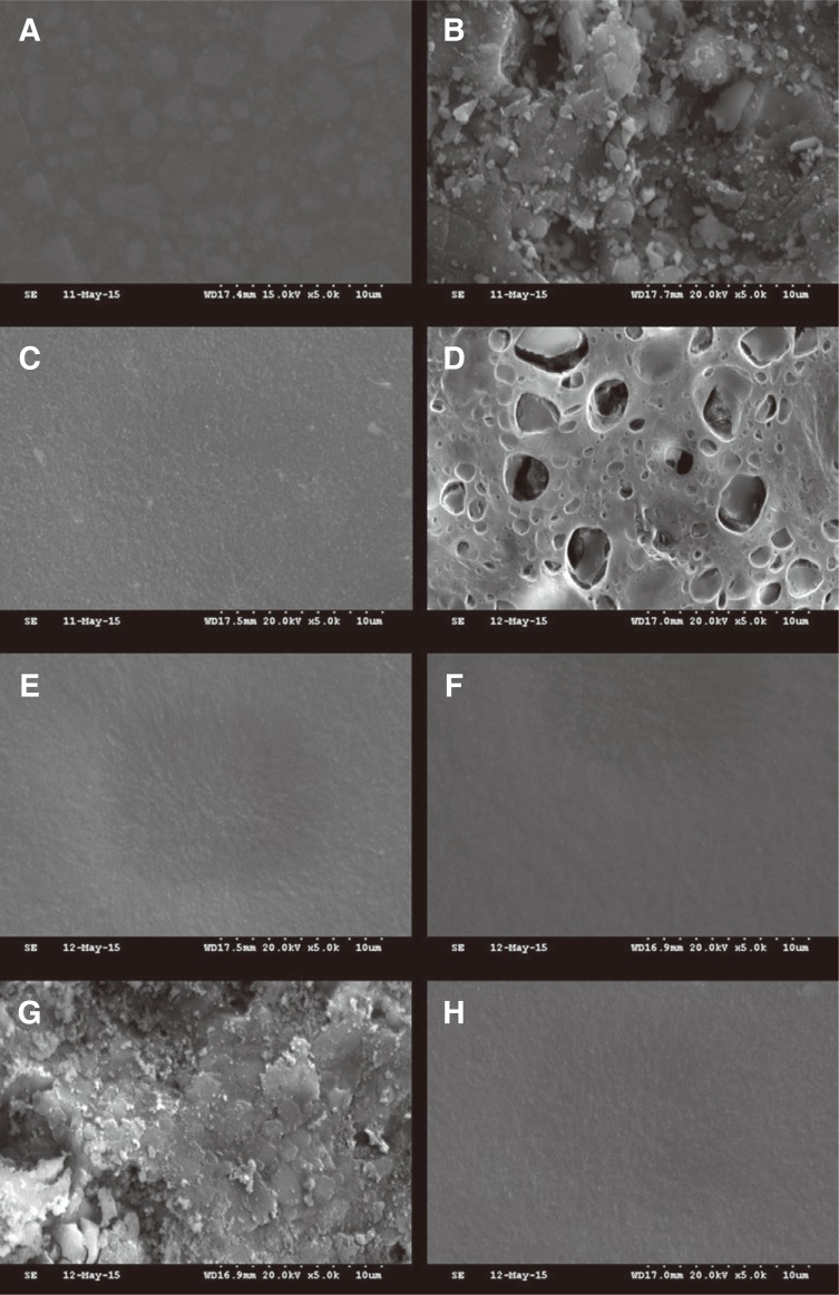
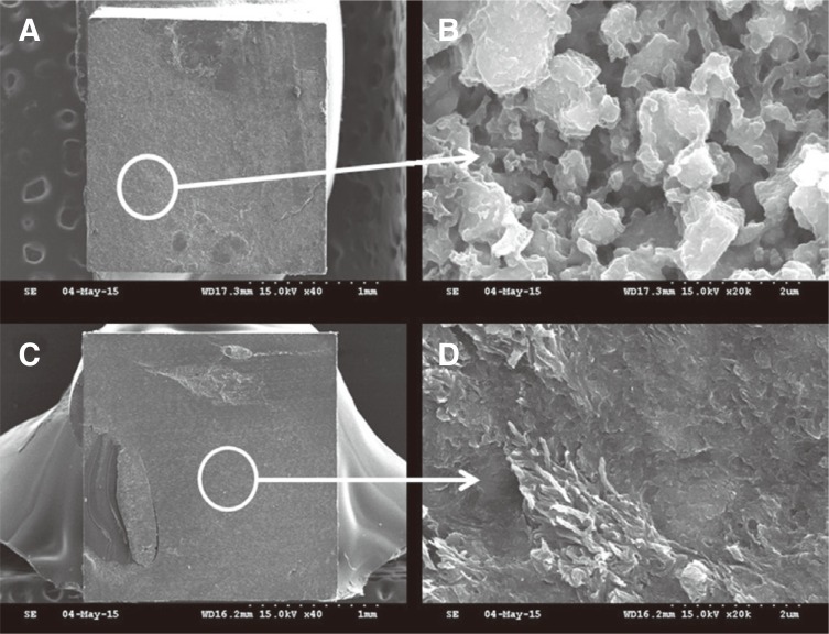
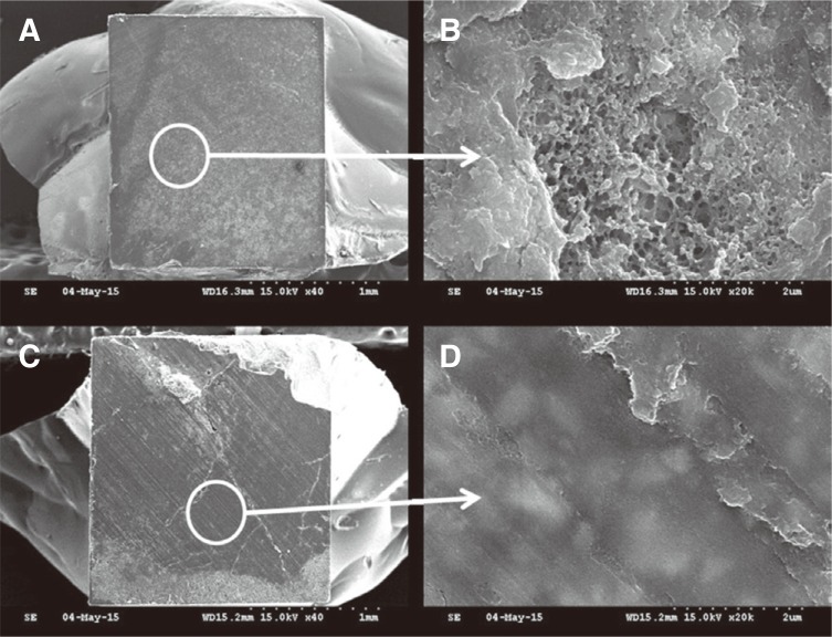
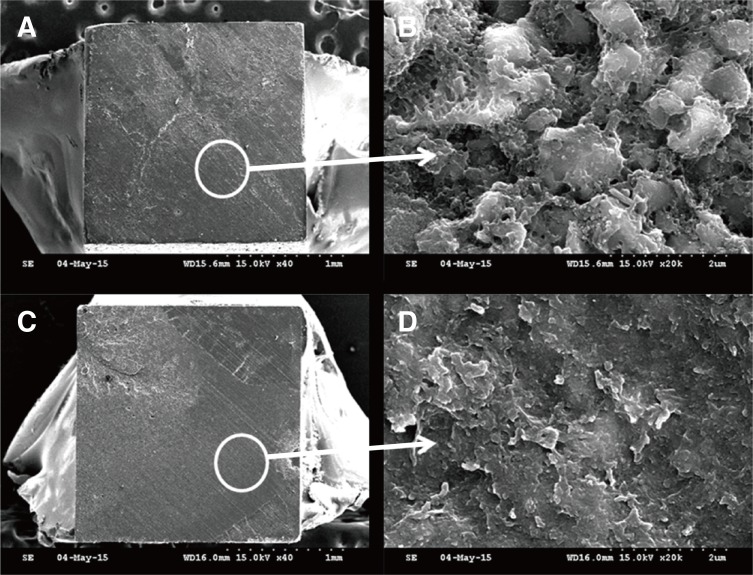
RNC onlays (Lava Ultimate) (n=30) were treated using air abrasion with and without a universal adhesive, or HF etching followed by a universal adhesive with and without a silane coupling agent, or tribological silica coating with and without a universal adhesive, and divided into 6 groups. Onlays were luted with resin cement to dentin surfaces. A microtensile bond strength test was performed and evaluated by one-way ANOVA and Tukey HSD test (α=.05). A nanoscratch test, field emission scanning electron microscopy, and energy dispersive X-ray spectroscopy were used for micromorphologic analysis (α=.05). The roughness and elemental proportion were evaluated by Kruskal-Wallis test and Mann-Whitney U test.
RESULTS
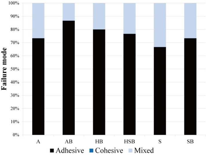
Tribological silica coating showed the highest roughness, followed by air abrasion and HF etching. After HF etching, the RNC surface presented a decrease in oxygen, silicon, and zirconium ratio with increasing carbon ratio. Air abrasion with universal adhesive showed the highest bond strength followed by tribological silica coating with universal adhesive. HF etching with universal adhesive showed the lowest bond strength.
CONCLUSION
An improved understanding of the effect of surface treatment of RNC could enhance the durability of resin bonding when used for indirect restorations. When using RNC for restoration, effective and systemic surface roughening methods and an appropriate adhesive are required.
Keyword
MeSH Terms
Figure
Cited by 1 articles
-
Surface deterioration of monolithic CAD/CAM restorative materials after artificial abrasive toothbrushing
Nazmiye Şen, Betül Tuncelli, Gültekin Göller
J Adv Prosthodont. 2018;10(4):271-278. doi: 10.4047/jap.2018.10.4.271.
Reference
-
1. Alt V, Hannig M, Wöstmann B, Balkenhol M. Fracture strength of temporary fixed partial dentures: CAD/CAM versus directly fabricated restorations. Dent Mater. 2011; 27:339–347. PMID: 21176946.
Article2. Schoenbaum TR. Dentistry in the digital age: an update. Dent Today. 2012; 31:108110112–113. PMID: 22413391.3. Poticny DJ, Klim J. CAD/CAM in-office technology: innovations after 25 years for predictable, esthetic outcomes. J Am Dent Assoc. 2010; 141:5S–9S. PMID: 20516108.4. Stawarczyk B, Ender A, Trottmann A, Özcan M, Fischer J, Hämmerle CH. Load-bearing capacity of CAD/CAM milled polymeric three-unit fixed dental prostheses: effect of aging regimens. Clin Oral Investig. 2012; 16:1669–1677.
Article5. Tinschert J, Zwez D, Marx R, Anusavice KJ. Structural reliability of alumina-, feldspar-, leucite-, mica- and zirconia-based ceramics. J Dent. 2000; 28:529–535. PMID: 10960757.
Article6. Stawarczyk B, Sener B, Trottmann A, Roos M, Ozcan M, Hämmerle CH. Discoloration of manually fabricated resins and industrially fabricated CAD/CAM blocks versus glass-ceramic: effect of storage media, duration, and subsequent polishing. Dent Mater J. 2012; 31:377–383. PMID: 22673470.7. Stawarczyk B, Özcan M, Trottmann A, Schmutz F, Roos M, Hämmerle C. Two-body wear rate of CAD/CAM resin blocks and their enamel antagonists. J Prosthet Dent. 2013; 109:325–332. PMID: 23684283.
Article8. Luthardt RG, Holzhüter MS, Rudolph H, Herold V, Walter MH. CAD/CAM-machining effects on Y-TZP zirconia. Dent Mater. 2004; 20:655–662. PMID: 15236940.
Article9. Lin WS, Ercoli C, Feng C, Morton D. The effect of core material, veneering porcelain, and fabrication technique on the biaxial flexural strength and weibull analysis of selected dental ceramics. J Prosthodont. 2012; 21:353–362. PMID: 22462639.
Article10. Addison O, Cao X, Sunnar P, Fleming GJ. Machining variability impacts on the strength of a 'chair-side' CAD-CAM ceramic. Dent Mater. 2012; 28:880–887. PMID: 22579103.
Article11. El Zohairy AA, De Gee AJ, Mohsen MM, Feilzer AJ. Microtensile bond strength testing of luting cements to prefabricated CAD/CAM ceramic and composite blocks. Dent Mater. 2003; 19:575–583. PMID: 12901980.
Article12. 3M ESPE. Lava ultimate technical product profile: 2011. Available at: http://multimedia.3m.com/mws/media/781394O/lava-ultimate-technical-product-profilegolbal.pdf.13. Chen C, Trindade FZ, de Jager N, Kleverlaan CJ, Feilzer AJ. The fracture resistance of a CAD/CAM Resin Nano Ceramic (RNC) and a CAD ceramic at different thicknesses. Dent Mater. 2014; 30:954–962. PMID: 25037897.
Article14. Joda T, Huber S, Bürki A, Zysset P, Brägger U. Influence of Abutment Design on Stiffness, Strength, and Failure of Implant-Supported Monolithic Resin Nano Ceramic (RNC) Crowns. Clin Implant Dent Relat Res. 2015; 17:1200–1207. PMID: 24629091.
Article15. Vargas MA, Bergeron C, Diaz-Arnold A. Cementing all-ceramic restorations: recommendations for success. J Am Dent Assoc. 2011; 142:20S–24S. PMID: 21454837.16. Spitznagel FA, Horvath SD, Guess PC, Blatz MB. Resin bond to indirect composite and new ceramic/polymer materials: a review of the literature. J Esthet Restor Dent. 2014; 26:382–393. PMID: 24754327.
Article17. el-Mowafy O. The use of resin cements in restorative dentistry to overcome retention problems. J Can Dent Assoc. 2001; 67:97–102. PMID: 11253298.18. Sorensen JA, Kang SK, Avera SP. Porcelain-composite interface microleakage with various porcelain surface treatments. Dent Mater. 1991; 7:118–123. PMID: 1718804.
Article19. Thompson JY, Stoner BR, Piascik JR, Smith R. Adhesion/cementation to zirconia and other non-silicate ceramics: where are we now? Dent Mater. 2011; 27:71–82. PMID: 21094526.
Article20. Semmelman JO, Kulp PR. Silane bonding porcelain teeth to acrylic. J Am Dent Assoc. 1968; 76:69–73. PMID: 5234516.
Article21. Jochen DG, Caputo AA. Composite resin repair of porcelain denture teeth. J Prosthet Dent. 1977; 38:673–679. PMID: 340655.
Article22. Ferrando JM, Graser GN, Tallents RH, Jarvis RH. Tensile strength and microleakage of porcelain repair materials. J Prosthet Dent. 1983; 50:44–50. PMID: 6135801.
Article23. Blatz MB, Sadan A, Kern M. Resin-ceramic bonding: a review of the literature. J Prosthet Dent. 2003; 89:268–274. PMID: 12644802.
Article24. Ozcan M. Evaluation of alternative intra-oral repair techniques for fractured ceramic-fused-to-metal restorations. J Oral Rehabil. 2003; 30:194–203. PMID: 12535148.25. Amaral R, Ozcan M, Bottino MA, Valandro LF. Microtensile bond strength of a resin cement to glass infiltrated zirconia-reinforced ceramic: the effect of surface conditioning. Dent Mater. 2006; 22:283–290. PMID: 16039705.
Article26. Ozcan M. The use of chairside silica coating for different dental applications: a clinical report. J Prosthet Dent. 2002; 87:469–472. PMID: 12070506.27. Bailey LF, Bennett RJ. DICOR surface treatments for enhanced bonding. J Dent Res. 1988; 67:925–931. PMID: 3049721.
Article28. Egilmez F, Ergun G, Cekic-Nagas I, Vallittu PK, Ozcan M, Lassila LV. Effect of surface modification on the bond strength between zirconia and resin cement. J Prosthodont. 2013; 22:529–536. PMID: 23551581.
Article29. Sorensen JA, Engelman MJ, Torres TJ, Avera SP. Shear bond strength of composite resin to porcelain. Int J Prosthodont. 1991; 4:17–23. PMID: 2012666.30. Chen JH, Matsumura H, Atsuta M. Effect of different etching periods on the bond strength of a composite resin to a machinable porcelain. J Dent. 1998; 26:53–58. PMID: 9479926.
Article31. Chen JH, Matsumura H, Atsuta M. Effect of etchant, etching period, and silane priming on bond strength to porcelain of composite resin. Oper Dent. 1998; 23:250–257. PMID: 9863446.32. Szep S, Gerhardt T, Gockel HW, Ruppel M, Metzeltin D, Heidemann D. In vitro dentinal surface reaction of 9.5% buffered hydrofluoric acid in repair of ceramic restorations: a scanning electron microscopic investigation. J Prosthet Dent. 2000; 83:668–674. PMID: 10842137.
Article33. Braga RR, Meira JB, Boaro LC, Xavier TA. Adhesion to tooth structure: a critical review of "macro" test methods. Dent Mater. 2010; 26:e38–e49. PMID: 20004960.
Article34. Scherrer SS, Cesar PF, Swain MV. Direct comparison of the bond strength results of the different test methods: a critical literature review. Dent Mater. 2010; 26:e78–e93. PMID: 20060160.
Article35. Papia E, Larsson C, du Toit M, Vult von Steyern P. Bonding between oxide ceramics and adhesive cement systems: a systematic review. J Biomed Mater Res B Appl Biomater. 2014; 102:395–413. PMID: 24123837.
Article36. Sano H, Shono T, Sonoda H, Takatsu T, Ciucchi B, Carvalho R, Pashley DH. Relationship between surface area for adhesion and tensile bond strength-evaluation of a micro-tensile bond test. Dent Mater. 1994; 10:236–240. PMID: 7664990.37. Pashley DH, Carvalho RM, Sano H, Nakajima M, Yoshiyama M, Shono Y, Fernandes CA, Tay F. The microtensile bond test: a review. J Adhes Dent. 1999; 1:299–309. PMID: 11725659.38. Blatz MB, Phark JH, Ozer F, Mante FK, Saleh N, Bergler M, Sadan A. In vitro comparative bond strength of contemporary self-adhesive resin cements to zirconium oxide ceramic with and without air-particle abrasion. Clin Oral Investig. 2010; 14:187–192.
Article39. Soares CJ, Soares PV, Pereira JC, Fonseca RB. Surface treatment protocols in the cementation process of ceramic and laboratory-processed composite restorations: a literature review. J Esthet Restor Dent. 2005; 17:224–235. PMID: 16231493.
Article40. Borges GA, Sophr AM, de Goes MF, Sobrinho LC, Chan DC. Effect of etching and airborne particle abrasion on the microstructure of different dental ceramics. J Prosthet Dent. 2003; 89:479–488. PMID: 12806326.
Article41. Swift EJ Jr, Brodeur C, Cvitko E, Pires JA. Treatment of composite surfaces for indirect bonding. Dent Mater. 1992; 8:193–196. PMID: 1521710.
Article42. Kern M, Thompson VP. Sandblasting and silica coating of a glass-infiltrated alumina ceramic: volume loss, morphology, and changes in the surface composition. J Prosthet Dent. 1994; 71:453–461. PMID: 8006839.
Article43. Hantsche H. Comparison of basic principles of the surface-specific analytical methods: AES/SAM, ESCA (XPS), SIMS, and ISS with X-ray microanalysis, and some applications in research and industry. Scanning. 1989; 11:257–280.
Article44. Pashley DH, Sano H, Ciucchi B, Yoshiyama M, Carvalho RM. Adhesion testing of dentin bonding agents: a review. Dent Mater. 1995; 11:117–125. PMID: 8621032.
Article45. Imamura GM, Reinhardt JW, Boyer DB, Swift EJ Jr. Enhancement of resin bonding to heat-cured composite resin. Oper Dent. 1996; 21:249–256. PMID: 9227119.46. Janda R, Roulet JF, Wulf M, Tiller HJ. A new adhesive technology for all-ceramics. Dent Mater. 2003; 19:567–573. PMID: 12837406.
Article47. Stawarczyk B, Krawczuk A, Ilie N. Tensile bond strength of resin composite repair in vitro using different surface preparation conditionings to an aged CAD/CAM resin nanoceramic. Clin Oral Investig. 2015; 19:299–308.
Article48. D'Arcangelo C, Vanini L. Effect of three surface treatments on the adhesive properties of indirect composite restorations. J Adhes Dent. 2007; 9:319–326. PMID: 17655072.
- Full Text Links
- Actions
-
Cited
- CITED
-
- Close
- Share
- Similar articles
-
- Repair bond strengths of non-aged and aged resin nanoceramics
- The effect of Er,Cr:YSGG irradiation on microtensile bond strength of composite resin restoration
- The etching effects and microtensile bond strength of total etching and self-etching adhesive system on unground enamel
- Bonding of a resin-modified glass ionomer cement to dentin using universal adhesives
- The Effect of Temporary Filling Materials on The Adhesion between Dentin Adhesive-coated Surface and Resin Inlay

