Melanocytic Nevus on the Rectal Mucosa Removed Using Endoscopic Submucosal Dissection
- Affiliations
-
- 1Division of Gastroenterology, Department of Internal Medicine, Korea University Guro Hospital, Korea University College of Medicine, Seoul, Korea. gi7pjj@korea.ac.kr
- KMID: 2348261
- DOI: http://doi.org/10.5946/ce.2015.126
Abstract
- Melanocytic nevus is the benign proliferation of melanocytes. The most common location of melanocytic nevus is the skin of the extremities; however, there are few case reports of melanocytic nevus at the rectal mucosa. No prior case of malignant melanoma from melanocytic nevus at the rectal mucosa has been reported; therefore, it is unclear whether resection should be performed or close observation is sufficient. However, the potential malignant transformation of melanocytic nevus should be considered, including melanocytic nevus on the rectum. Melanocytic nevus of the skin can be removed by surgical excision; however, due to rare incidence on the mucosa of the gastrointestinal tract, the optimal treatment for rectal melanocytic nevus remains controversial. Here, we report the first case of melanocytic nevus on the rectal mucosa that was removed by endoscopic submucosal dissection. This case report provides useful information about the optimal management of rectal melanocytic nevus.
MeSH Terms
Figure
-
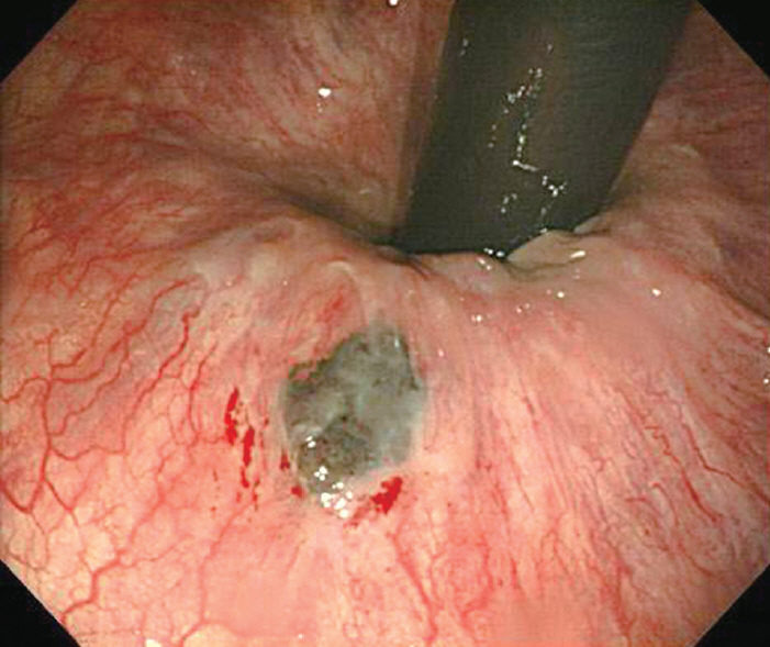
Fig. 1. Endoscopic finding of rectal melanocytic nevus. A 1.0×0.8 cm hyperpigmented and slightly
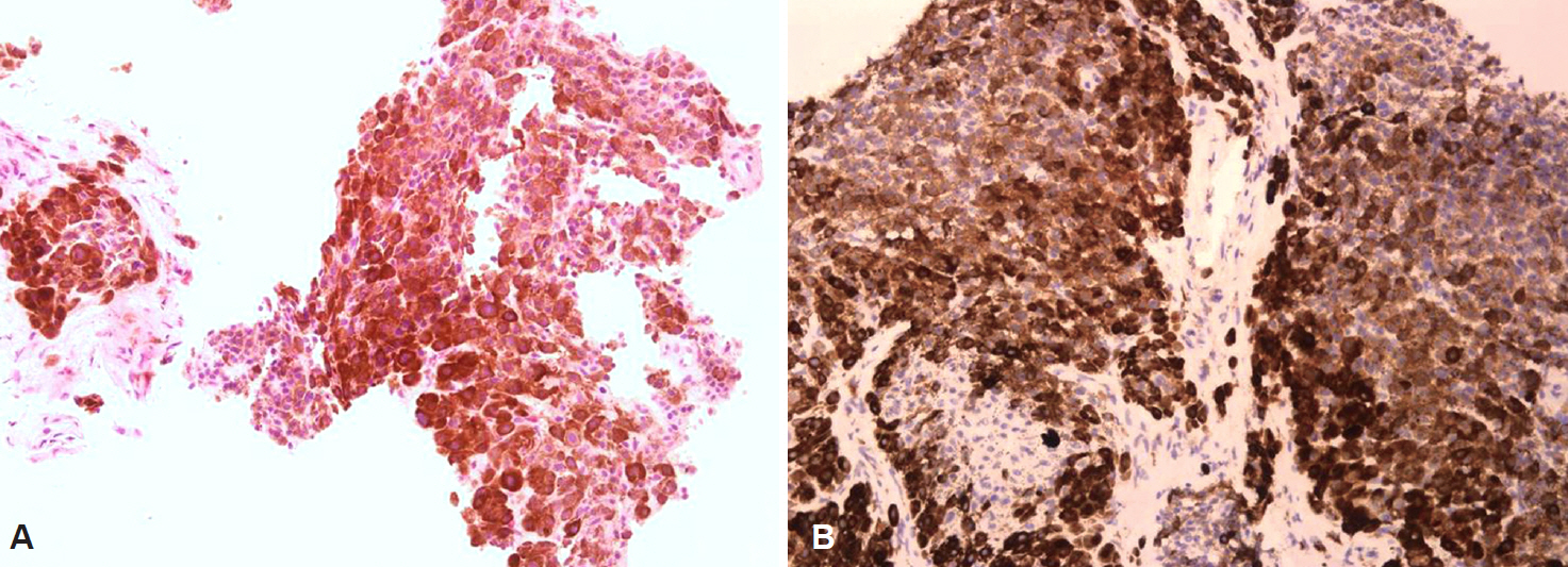
Fig. 2. Histopathological findings of endoscopic biopsy tissue. (A) The sample is composed of many melanocytes and brown pigmentation without malignant cells (H&E stain, ×40). (B) The Ki-67 labeling index of the specimen is <1% (×200).
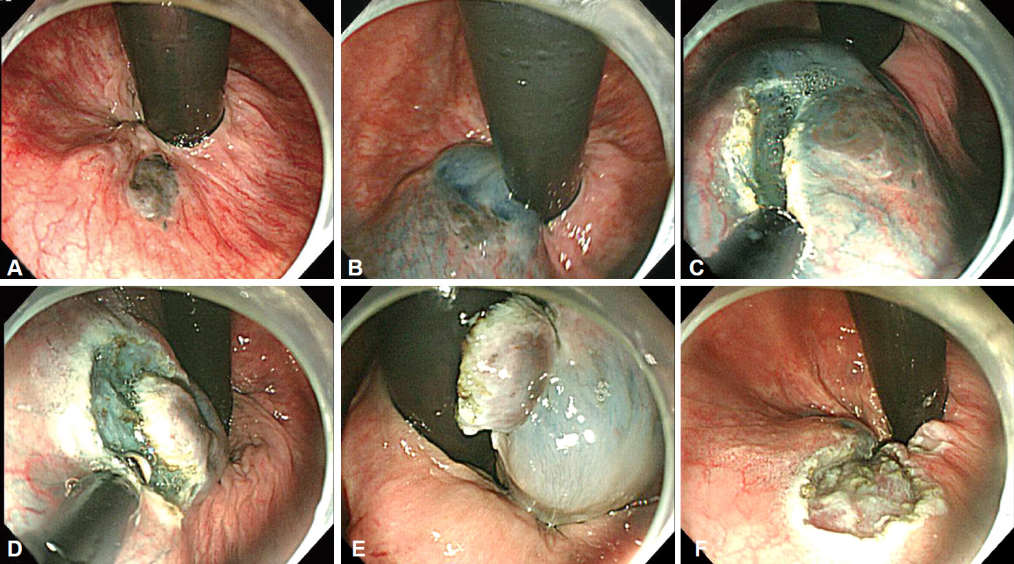
Fig. 3. Endoscopic submucosal dissection of melanocytic nevus of the rectum. (A) A rectal melanocytic nevus is visible on the far-distal rectum. (B) A submucosal injection is performed using epinephrine-indigo carmine-saline solution. (C) A mucosal incision around the lesion is made using a needle knife. (D, E) The submucosal dissection is performed using an insulated-tip knife. (F) The rectal melanocytic nevus is completely removed without major complications.
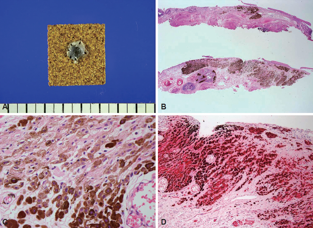
Fig. 4. Histopathological findings of the resected specimen. (A) The gross findings of the specimen indicate that the lesion is 0.7×0.7 cm. (B) The nevus involves the submucosa of the anorectal junction (H&E stain, ×12.5). (C) The resected lesion is composed of many melanocytes without malignant cells. Almost all of the cells include brown pigmentation (H&E stain, ×400). (D) The lesion is positive for S-100 (×100).
Reference
-
1. Gilchrest BA, Eller MS, Geller AC, Yaar M. The pathogenesis of melanoma induced by ultraviolet radiation. N Engl J Med. 1999; 340:1341–1348.
Article2. Chaudru V, Chompret A, Bressac-de Paillerets B, Spatz A, Avril MF, Demenais F. Influence of genes, nevi, and sun sensitivity on melanoma risk in a family sample unselected by family history and in melanoma-prone families. J Natl Cancer Inst. 2004; 96:785–795.
Article3. Purdue MP, From L, Armstrong BK, et al. Etiologic and other factors predicting nevus-associated cutaneous malignant melanoma. Cancer Epidemiol Biomarkers Prev. 2005; 14:2015–2022.
Article4. Bataille V, Bishop JA, Sasieni P, et al. Risk of cutaneous melanoma in relation to the numbers, types and sites of naevi: a case-control study. Br J Cancer. 1996; 73:1605–1611.
Article5. Lynch HT, Frichot BC 3rd, Lynch JF. Familial atypical multiple mole-melanoma syndrome. J Med Genet. 1978; 15:352–356.
Article6. Harrison SL, MacLennan R, Speare R, Wronski I. Sun exposure and melanocytic naevi in young Australian children. Lancet. 1994; 344:1529–1532.
Article7. Patrick RJ, Fenske NA, Messina JL. Primary mucosal melanoma. J Am Acad Dermatol. 2007; 56:828–834.
Article8. Khan M, Bucher N, Elhassan A, et al. Primary anorectal melanoma. Case Rep Oncol. 2014; 7:164–170.
Article9. Mason JK, Helwig EB. Ano-rectal melanoma. Cancer. 1966; 19:39–50.
Article
- Full Text Links
-
- Actions
-
Cited
- CITED
-
- Close
- Share
-
- Similar articles
-
- A Case of a Compound Nevus That Developed as Papillomatous Melanocytic Nevus
- Three Cases of Malignant Melanoma Possibly Arising in a Long Standing Melanocytic Nevus
- A Case of Congenital Melanocytic Nevus Combined with an Epidermal Cyst
- Alopecia Associated with Underlying Congenital Melanocytic Nevus
- Endoscopic treatment for rectal neuroendocrine tumor: which method is better?
