MRI Findings of Synovial Lipoma Arborescens of the Hip: A Case Report
- Affiliations
-
- 1Department of Radiology, Myoungji Hospital, Kwandong University College of Medicine, Korea.
- 2Department of Radiology, Shinchon Severance Hospital, Yonsei University School of Medicine, Korea.
- 3Department of Orthopedic Surgery, Myoungji Hospital, Kwandong University College of Medicine, Korea. wonnypia@kwandong.ac.kr
- 4Department of Pathology, Myoungji Hospital, Kwandong University College of Medicine, Korea.
Abstract
- Lipoma arborescens is a rare intra-articular lesion consisting of villous lipomatous proliferation of the synovium that tends to occur in the knee joint, especially in the suprapatellar pouch. The lesion has been observed in other locations, including the glenohumeral joint, subdeltoid bursa, hip, and elbow. We report a case of a lipoma arborescens of a unilateral hip joint in a 34-year-old man. MRI and a whole body bone scan were performed. In addition, a surgical synovectomy was performed and microscopic examination of the tumor revealed multiple variable-sized villous masses composed of mature fat tissue and lined by synovial cells which confirmed the diagnosis of lipoma arborescens.
MeSH Terms
Figure
-

Fig. 1 Plain radiograph reveals joint space narrowing and erosive changes in the inferomedial portion of the right femoral head, but no gross calcification. Extrinsic indentation in the medial aspect is also noted.

Fig. 2 Axial T1-weighted (A) and T2-weighted (B) MR images of the right hip show the frond-like villous lipomatous proliferation of the synovium with signal corresponding to the fat (arrow). A coronal T2-weighted fat-suppressed MR image (C) shows decreased signal intensity of synovial proliferation identical to that of subcutaneous fat (arrow).
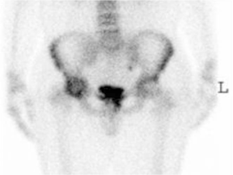
Fig. 3 A whole body bone scan (30 mci of Tc-99m MDP) shows increased uptake in the right hip joint.
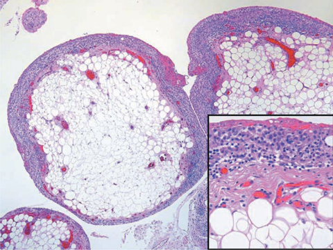
Fig. 4 Microscopic examination of the tumor reveals villous masses composed of mature adipocytes lined by synovial cells (×40, inlet ×200).
Reference
-
1. Feller JF, Rishi M, Hughes EC. Lipoma arborescens of the knee: MR demonstration. AJR Am J Roentgenol. 1994; 163:162–164.2. Ryu KN, Jaovisidha S, Schweitzer M, Motta AO, Resnick D. MR imaging of lipoma arborescens of the knee joint. AJR Am J Roentgenol. 1996; 176:1229–1232.3. Bejia I, Younes M, Moussa A, Said M, Touzi M, Bergaoui N. Lipoma arborescens affecting multiple joints. Skeletal Radiol. 2005; 34:536–538.4. Wolf RS, Zoys GN, Saldivar VA, Williams RP. Lipoma arborescens of the hip. Am J Orthop. 2002; 31:276–276.5. Noel ER, Tebib JG, Dumontet C, Colson F, Carret JP, Vauzelle JL, et al. Synovial lipoma arborescens of the hip. Clin Rheumatol. 1987; 6:92–96.6. Martin S, Hernandez L, Romero J, Lafuente J, Poza AI, Ruiz P, et al. Diagnostic imaging of lipoma arborescens. Skeletal Radiol. 1998; 27:325–329.7. Sola JB, Wright RW. Arthroscopic treatment for lipoma arborescens of the knee. J Bone Joint Surg Am. 1998; 80:99–103.8. Edamitsu S, Mizuta H, Kubota K, Matsukawa A. Lipoma arborescens with hemarthrosis of the knee: a case report. Acta Orthop Scand. 1993; 64:601–602.9. Yan CH, Wong JW, Yip DK. Bilateral knee lipoma arborescens: a case report. J Orthop Surg. 2008; 16:107–110.10. Hallel T, Lew S, Bansal M. Villous lipomatous proliferation of the synovial membrane (lipoma arborescens). J Bone Joint Surg Am. 1988; 70:264–270.
- Full Text Links
-
- Actions
-
Cited
- CITED
-
- Close
- Share
-
- Similar articles
-
- Lipoma Arborescens in the Hip Joint: A Case Report
- The Arthroscopic Treatment of Lipoma Arborescens of Knee: A Case Report
- Arthroscopic Treatment of the Lipoma Arborescens in All Compartments of the Knee: A Case Report
- Lipoma Arborescens in the Knee Joint: A Case Report
- Tenosynovial Bilateral Lipoma Arborescens of the Ankle in Adults
