Rare Lung Manifestation of Multifocal Micronodular Pneumocyte Hyperplasia in a Teenage Girl with Tuberous Sclerosis Complex
- Affiliations
-
- 1Department of Radiology, Kangwon National University Hospital, Chuncheon, Korea. hk2005.yoon@gmail.com
- 2Department of Pathology, Kangwon National University Hospital, Chuncheon, Korea.
- KMID: 2344803
- DOI: http://doi.org/10.3348/jksr.2016.75.2.133
Abstract
- Multifocal micronodular pneumocyte hyperplasia (MMPH) is a relatively rare pulmonary disorder that can be associated with tuberous sclerosis complex (TSC). It has been rarely reported in children or adolescents. MMPH is a hamartomatous process of the lung with multiple small nodules, composed of type II pneumocytes. Plain radiography and chest CT in MMPH may demonstrate numerous small nodules measuring 1-10 mm in diameters, distributed randomly throughout both lungs. If MMPH is an initial presentation of TSC, and unless we are familiar with this lung manifestation of TSC, radiologic findings can mimic miliary tuberculosis or metastatic disease. We report a teenage girl with TSC and histologically confirmed MMPH which mimicked miliary tuberculosis at the initial presentation.
MeSH Terms
Figure
-
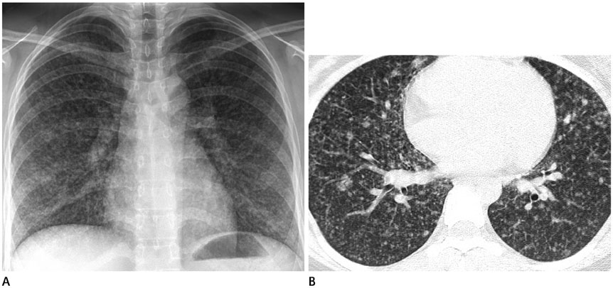
Fig. 1 Multifocal micronodular pneumocyte hyperplasia with tuberous sclerosis complex in a 15-year old girl. A. Initial chest radiograph for student screening shows numerous fine nodular opacities evenly scattered throughout both lungs. B. High resolution chest CT shows nodules and multiple, randomly distributed, 3–12 mm nodules. No cysts are seen.
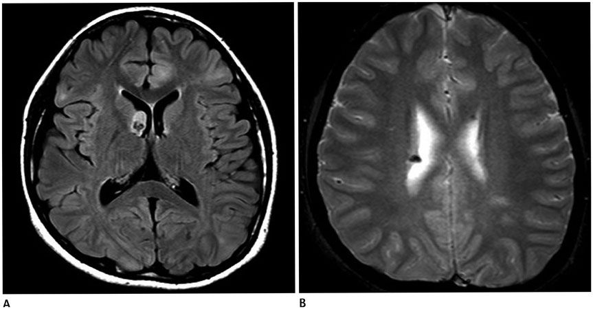
Fig. 2 Brain MR imaging of multifocal micronodular pneumocyte hyperplasia with tuberous sclerosis complex in a 15-year old girl. A. Axial FLAIR image shows a subependymal nodule near the right foramen Monro and multiple high signal intensities at the cerebral cortex and subcortical white matter suggesting cortical and subcortical tubers. B. Axial gradient echo image shows calcified subependymal nodules along the lateral ventricular margins. FLAIR = fluid attenuated inversion recovery, MR = magnetic resonance
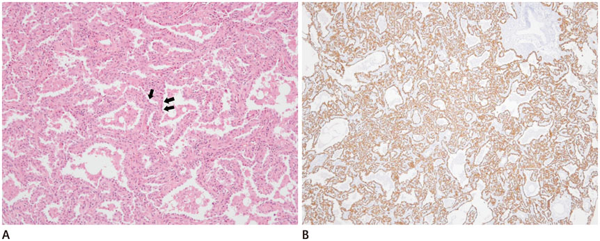
Fig. 3 Microscopic findings of VATS biopsy specimens of multifocal micronodular pneumocyte hyperplaisa with tuberous sclerosis complex in a 15-year old girl. A. Close-up view of the nodule show increased septal thickness and pneumocyte hyperplasia (arrows) (hematoxylin and eosin stain, × 100). B. Immunohistochemically, most of the proliferating epithelial cells of the lesion are positive for pan-cytokeratin (brown color) (pan-cytokeratin stain, × 40). VATS = video-assisted thoracoscopic surgery
Reference
-
1. Tee AR, Manning BD, Roux PP, Cantley LC, Blenis J. Tuberous sclerosis complex gene products, tuberin and hamartin, control mTOR signaling by acting as a GTPase-activating protein complex toward Rheb. Curr Biol. 2003; 13:1259–1268.2. Maruyama H, Ohbayashi C, Hino O, Tsutsumi M, Konishi Y. Pathogenesis of multifocal micronodular pneumocyte hyperplasia and lymphangioleiomyomatosis in tuberous sclerosis and association with tuberous sclerosis genes TSC1 and TSC2. Pathol Int. 2001; 51:585–594.3. Franz DN, Brody A, Meyer C, Leonard J, Chuck G, Dabora S, et al. Mutational and radiographic analysis of pulmonary disease consistent with lymphangioleiomyomatosis and micronodular pneumocyte hyperplasia in women with tuberous sclerosis. Am J Respir Crit Care Med. 2001; 164:661–668.4. Kobashi Y, Sugiu T, Mouri K, Irei T, Nakata M, Oka M. Clinicopathological analysis of multifocal micronodular pneumocyte hyperplasia associated with tuberous sclerosis in Japan. Respirology. 2008; 13:1076–1081.5. Behnes CL, Schütze G, Engelke C, Bremmer F, Gunawan B, Radzun HJ, et al. 13-year-old tuberous sclerosis patient with renal cell carcinoma associated with multiple renal angiomyolipomas developing multifocal micronodular pneumocyte hyperplasia. BMC Clin Pathol. 2013; 13:4.6. Northrup H, Krueger DA. International Tuberous Sclerosis Complex Consensus Group. Tuberous sclerosis complex diagnostic criteria update: recommendations of the 2012 Iinternational Tuberous Sclerosis Complex Consensus Conference. Pediatr Neurol. 2013; 49:243–254.7. Hong SH, Im JG, Lee JS, Song JW, Lee HJ, Yeon KM. High resolution CT findings of miliary tuberculosis. J Comput Assist Tomogr. 1998; 22:220–224.8. Webb WR, Muller NL, Naidich DP. High-resolution CT of the lung. Philadelphia: Lippincott Williams & Wilkins;2014. p. 368–381.9. Kushihashi T, Munechika H, Ri K, Kubota H, Ukisu R, Satoh S, et al. Bronchioloalveolar adenoma of the lung: CT-pathologic correlation. Radiology. 1994; 193:789–793.10. Fujitaka K, Isobe T, Oguri T, Yamasaki M, Miyazaki M, Kohno N, et al. A case of micronodular pneumocyte hyperplasia diagnosed through lung biopsy using thoracoscopy. Respiration. 2002; 69:277–279.
- Full Text Links
-
- Actions
-
Cited
- CITED
-
- Close
- Share
-
- Similar articles
-
- Pulmonary Lymphangioleiomyomatosis and Micronodular Pneumocyte Hyperplasia associated with Tuberous Sclerosis: A Case Report
- A Case of Multiple Micronodular Pneumocyte Hyperplasia of the Lung in a Man with Tuberous Sclerosis
- Concurrent renal and hepatic angiomyolipoma with pulmonary involvement in two patients with tuberous sclerosis
- A case of Renal Angiomyolipoma in a Child without Evidence of Tuberous Sclerosis
- A case of bilateral renal angiomyolipoma associated with tuberous sclerosis
