J Korean Soc Radiol.
2011 Nov;65(5):473-477.
Oncocytic Schneiderian Papilloma Presenting as an Intensely Hypermetabolic Lesion of the Maxillary Sinus on 18F-Fluorodeoxyglucose Positron Emission Tomography/CT: A Case Report and Literature Review
- Affiliations
-
- 1Department of Radiology, Dongsan Medical Center, Keimyung University School of Medicine, Daegu, Korea. sklee@dsmc.or.kr
- 2Department of Pathology, Dongsan Medical Center, Keimyung University School of Medicine, Daegu, Korea.
Abstract
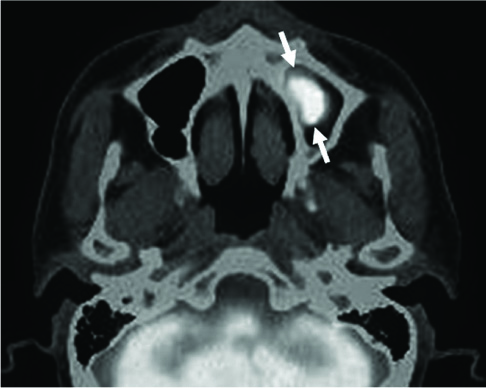
- A 54-year-old man presented with an incidentally identified intensely hypermetabolic lesion (SUVmax: 22.2 g/mL) in the left maxillary sinus on 18F-fluorodeoxyglucose positron emission tomography/computed tomography (18F-FDG PET/CT) performed for cancer screening. The mass was well circumscribed and showed solid enhancement on contrast-enhanced CT. Histological examination of the mass was consistent with oncocytic schneiderian papilloma. It is of prime importance to recognize that a sinonasal lesion with intense hypermetabolism on 18F-FDG PET/CT does not necessarily signify malignancy. Oncocytic schneiderian papilloma should be included in the differential diagnosis of an intensely hypermetabolic and solidly enhancing mass of the nasal cavities or paranasal sinuses.
MeSH Terms
Figure
Reference
-
1. Hyams VJ. Papillomas of the nasal cavity and paranasal sinuses. A clinicopathological study of 315 cases. Ann Otol Rhinol Laryngol. 1971; 80:192–206.2. Barnes L, Bedetti C. Oncocytic Schneiderian papilloma: a reappraisal of cylindrical cell papilloma of the sinonasal tract. Hum Pathol. 1984; 15:344–351.3. Kapadia SB, Barnes L, Pelzman K, Mirani N, Heffner DK, Bedetti C. Carcinoma ex oncocytic Schneiderian (cylindrical cell) papilloma. Am J Otolaryngol. 1993; 14:332–338.4. Cohen EG, Baredes S, Zuckier LS, Mirani NM, Liu Y, Ghesani NV. 18F-FDG PET evaluation of sinonasal papilloma. AJR Am J Roentgenol. 2009; 193:214–217.5. Lin FY, Genden EM, Lawson WL, Som P, Kostakoglu L. High uptake in schneiderian papillomas of the maxillary sinus on positron-emission tomography using fluorodeoxyglucose. AJNR Am J Neuroradiol. 2009; 30:428–430.6. Lin E. FDG uptake in a benign paranasal sinus schneiderian papilloma. Clin Nucl Med. 2007; 32:338–339.7. Udaka T, Shiomori T, Nagatani G, Hisaoka M, Kakeda S, Korogi Y, et al. Oncocytic schneiderian papilloma confined to the sphenoid sinus detected by FDG-PET. Rhinology. 2007; 45:89–92.8. Cook GJ. Pitfalls in PET/CT interpretation. Q J Nucl Med Mol Imaging. 2007; 51:235–243.9. Gorospe L, Raman S, Echeveste J, Avril N, Herrero Y, Herna Ndez S. Whole-body PET/CT: spectrum of physiological variants, artifacts and interpretative pitfalls in cancer patients. Nucl Med Commun. 2005; 26:671–687.10. Ninomiya H, Oriuchi N, Kahn N, Higuchi T, Endo K, Takahashi K, et al. Diagnosis of tumor in the nasal cavity and paranasal sinuses with [11C]choline PET: comparative study with 2-[18F]fluoro-2-deoxy-D-glucose (FDG) PET. Ann Nucl Med. 2004; 18:29–34.11. Shojaku H, Fujisaka M, Yasumura S, Ishida M, Tsubota M, Nishida H, et al. Positron emission tomography for predicting malignancy of sinonasal inverted papilloma. Clin Nucl Med. 2007; 32:275–278.12. Kim DJ, Chung JJ, Ryu YH, Hong SW, Yu JS, Kim JH. Adrenocortical oncocytoma displaying intense activity on 18F-FDG-PET: a case report and a literature review. Ann Nucl Med. 2008; 22:821–824.13. Noji T, Kondo S, Hirano S, Ambo Y, Tanaka E, Katoh C, et al. Intraductal oncocytic papillary neoplasm of the pancreas shows strong positivity on FDG-PET. Int J Gastrointest Cancer. 2002; 32:43–46.14. Lee SK, Rho BH, Woo SK. Hürthle cell neoplasm: correlation of gray-scale and power Doppler sonographic findings with gross pathology. J Clin Ultrasound. 2010; 38:169–176.15. Tallini G. Oncocytic tumours. Virchows Arch. 1998; 433:5–12.16. Brown RS, Goodman TM, Zasadny KR, Greenson JK, Wahl RL. Expression of hexokinase II and Glut-1 in untreated human breast cancer. Nucl Med Biol. 2002; 29:443–453.17. Mamede M, Higashi T, Kitaichi M, Ishizu K, Ishimori T, Nakamoto Y, et al. [18F]FDG uptake and PCNA, Glut-1, and Hexokinase-II expressions in cancers and inflammatory lesions of the lung. Neoplasia. 2005; 7:369–379.18. Kadowaki T, Yano S, Araki K, Tokushima T, Morioka N. A case of pulmonary typical carcinoid with an extensive oncocytic component showing intense uptake of FDG. Thorax. 2011; 66:361–362.
- Full Text Links
- Actions
-
Cited
- CITED
-
- Close
- Share
- Similar articles
-
- A Case of Oncocytic Schneiderian Papilloma in the Maxillary Sinus
- A Case of Paranasal Sinus Papilloma with Increased FDG Uptake
- CT, MRI, and 18F-Fluorodeoxyglucose Positron Emission Tomography/CT Features of Primary Mucosal Melanoma Involving the Lacrimal Drainage Apparatus: A Case Report
- Clinically Occult Diffuse Large B-Cell Lymphoma of the Middle Turbinate Identified Using ¹â¸F-Fluorodeoxyglucose Positron Emission Tomography/Computed Tomography: A Case Report
- Positron Emission Tomography-CT, CT, and MR Imaging Findings of Tumor-Mimicking Organized Hematoma in the Maxillary Sinus: Two Case Reports


