Clinically Occult Diffuse Large B-Cell Lymphoma of the Middle Turbinate Identified Using ¹â¸F-Fluorodeoxyglucose Positron Emission Tomography/Computed Tomography: A Case Report
- Affiliations
-
- 1Department of Radiology, Keimyung University, Dongsan Hospital, Daegu, Korea. sklee@dsmc.or.kr
- 2Department of Pathology, Keimyung University, Dongsan Hospital, Daegu, Korea.
- KMID: 2454034
- DOI: http://doi.org/10.3348/jksr.2019.80.3.548
Abstract
- We report a case of clinically occult diffuse large B-cell lymphoma (DLBCL) of the middle turbinate (MT) identified by ¹â¸F-fluorodeoxyglucose positron emission tomography/computed tomography (¹â¸F-FDG PET/CT) in a 71-year-old man along with imaging findings. DLBCL was presented with a hypermetabolic right MT [maximum standardized uptake values (SUV(max)) = 8.8 gm/dL] on ¹â¸F-FDG PET/CT, while rhinologic examination was normal. CT showed nothing but slightly more intense enhancement of the right MT compared with the opposite side. The disease progressed during next 7 months until follow-up CT demonstrated solidly enhancing mass occupying entire right nasal cavity which was intensely hypermetabolic (SUV(max) = 12.8 gm/dL). Surgical biopsy confirmed the diagnosis. Follow-up CT and 18F-FDG PET/CT performed after chemotherapy demonstrated complete resolution of DLBCL of the right nasal cavity including the right MT. This is thought to be the first case report in the literature concerning clinically occult DLBCL presenting as a hypermetabolic MT on ¹â¸F-FDG PET/CT.
MeSH Terms
Figure
-
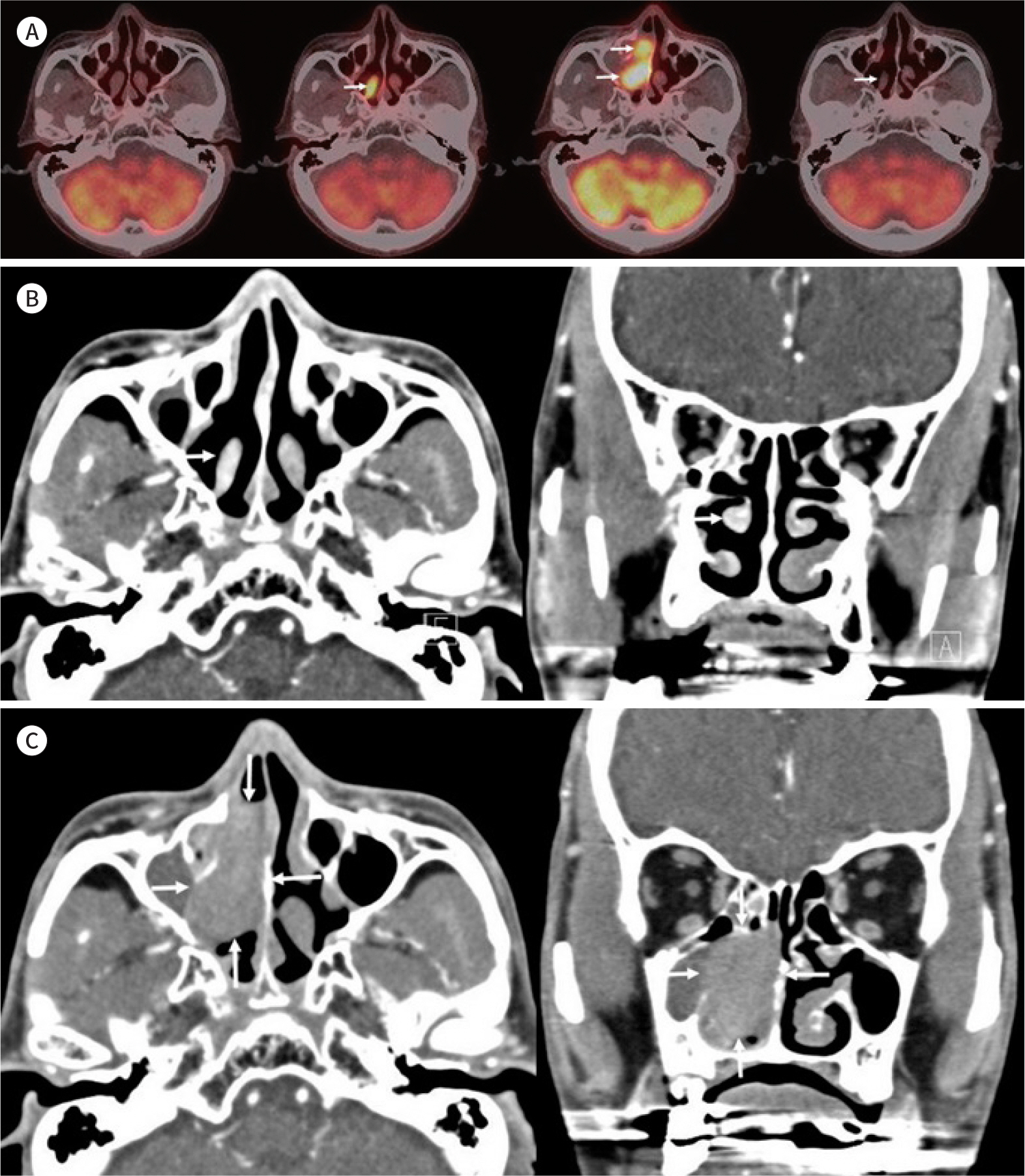
Fig. 1. A 71-year-old man with DLBCL of the MT detected using18F-FDG PET/CT that was performed for metastasis work-up of HCC. A. 18F-FDG PET/CT performed 14 months prior when he had undergone partial hepatectomy reveals no abnormal uptake in the sinonasal area (first image).18F-FDG PET/CT performed for metastasis work-up of HCC demonstrates hypermetabolic right MT (SUVmax = 8.8 gm/dL) (arrow) (second image). No abnormal uptake is noted at other sinonasal areas.18F-FDG PET/CT obtained 7 months thereafter shows an intensely hypermet-abolic mass (SUVmax = 12.8 gm/dL) (arrows) occupying the right nasal cavity (third image).18F-FDG PET/CTafter R-CHOP chemotherapy shows complete resolution of DLBCL of the right nasal cavity with normal appearing MT (arrow) (fourth image). B. Contrast-enhanced axial (first image) and coronal (second image) CT for further evaluation of hypermeta-bolic MT on18F-FDG PET/CT show morphologically normal right MT with slightly more intense enhancement compared to that on the opposite side (arrows). C. Follow-up contrast-enhanced axial CT (first image) obtained 7 months thereafter shows a mass with moderate enhancement in the right nasal cavity (arrows). Bony erosion and remodeling of the medial wall of the right maxillary sinus, right middle and inferior turbinates, and right ethmoid sinus are also noted on the coronal CT (arrows) (second image). CT = computed tomography, DLBCL = diffuse large B-cell lymphoma,18F-FDG =18F-fluorodeoxyglucose, HCC = hepatocellular carcinoma, MT = middle turbinate, PET = positron emission tomography, R-CHOP = cyclophosphamide, doxorubicin, vincristine, prednisolone, rituximab, SUVmax = maximum standardized uptake values
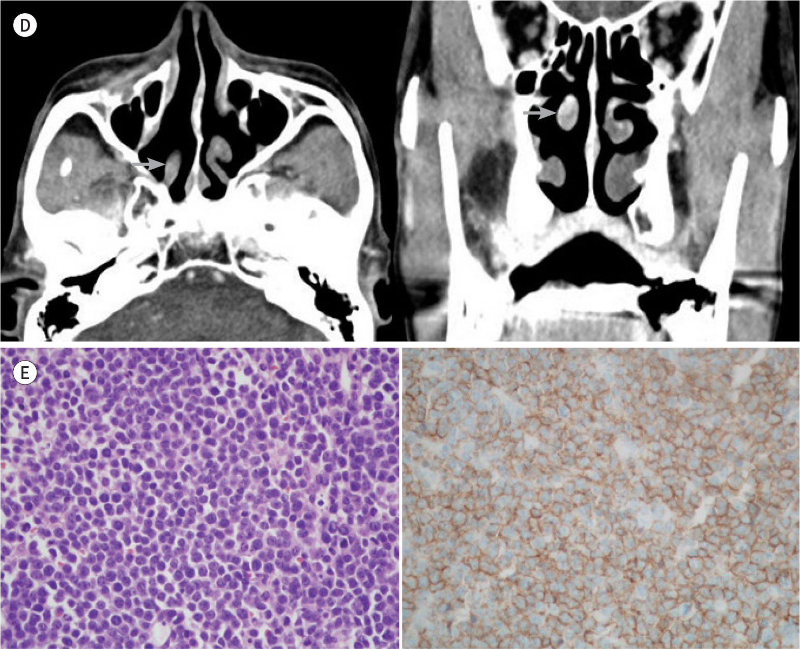
Fig. 1. A 71-year-old man with DLBCL of the MT detected using18F-FDG PET/CT that was performed for metastasis work-up of HCC. D. Follow-up axial (first image) and coronal (second image) CT after R-CHOP chemotherapy shows complete resolution of the mass of the right nasal cavity with normal-appearing MT (arrows). E. Histologic findings of the specimen obtained by surgical biopsy of the mass of the right nasal cavity demonstrates monotonous proliferation of medium to large lymphoid cells (first image; hematoxylin and eosin stain, × 200) with positivity for CD20 (second image; immunostaining, × 200). CT = computed tomography, DLBCL = diffuse large B-cell lymphoma,18F-FDG =18F-fluorodeoxyglucose, HCC = hepatocellular carcinoma, MT = middle turbinate, PET = positron emission tomography, R-CHOP = cyclophosphamide, doxorubicin, vincristine, prednisolone, rituximab
Reference
-
References
1. Woo JS, Kim JM, Lee SH, Chae SW, Hwang SJ, Lee HM. Clinical analysis of extranodal non-Hodgkin's lymphoma in the sinonasal tract.Eur Arch Otorhinolaryngol. 2004; 261:197–201.2. Oprea C, Cainap C, Azoulay R, Assaf E, Jabbour E, Koscielny S, et al. Primary diffuse large B-cell non-Hodgkin lymphoma of the paranasal sinuses: a report of 14 cases.Br J Haematol. 2005; 131:468–471.3. Yasumoto M, Taura S, Shibuya H, Honda M. Primary malignant lymphoma of the maxillary sinus: CT and MRI. Neuroradiology. 2000; 42:285–289.
Article4. Jang YM, Lee JH, Lee DH, Choi CG, Suh DC, Lee HK, et al. Imaging features of nasal-type natural killler/t-cell lymphomas: frequent involvement of skin and subcutaneous tissue.J Korean Radiol Soc. 2006; 54:161–165.5. Wang Q, Xiao S, Qin Y. Tumors originated from the inferior nasal turbinates: clinical features in 34 patients. Lin Chung Er Bi Yan Hou Tou Jing Wai Ke Za Zhi. 2014; 28:1050–1052.6. Lee JY, Jang YD, Kim HK. The primary role of the otolaryngologist in managing pediatric sinonasal malignancies: an extranodal NK/T-cell lymphoma originating from the inferior turbinate mucosa of the nasal cavity. J Pediatr Hematol Oncol. 2008; 30:401–404.
Article7. Lingan MB, Roasa FV. Intravascular lymphoma of the inferior turbinate: an unusual rhinologic presentation of a rare neoplasm.Philipp J Otolaryngol Head Neck Surg. 2007; 22:24–26.8. Jeong YJ, Sohn MH, Lim ST, Kim DW, Jeong HJ, Jang KY, et al. Osteoblastoma in the nasal cavity: F-18 FDG PET/CT and Tc-99m MDP 3-phase bone scan findings with pathologic correlation.Clin Nucl Med. 2011; 36:214–217.9. Arens AI, Verbist BM, Hendrickx BW, De Geus-Oei LF, Oyen WJ. False positive 18F-FDG PET/CT due to inflamed concha bullosa. Clin Nucl Med. 2012; 37:509–510.
Article10. Purohit BS, Ailianou A, Dulguerov N, Becker CD, Ratib O, Becker M. FDG-PET/CT pitfalls in oncological head and neck imaging. Insights Imaging. 2014; 5:585–602.
Article
- Full Text Links
-
- Actions
-
Cited
- CITED
-
- Close
- Share
-
- Similar articles
-
- Fluorine-18 Fluorodeoxyglucose Positron Emission Tomography/Computed Tomography Findings of Post Traumatic Lymphangioma in a Young Adult Male
- â¶â¸Gallium-Arginine-Glycine-Aspartic Acid and ¹â¸F-Fluorodeoxyglucose Positron Emission Tomography/Computed Tomography in Chondroblastic Osteosarcoma of the Skull
- A Case of Fluorine-18-Fluorodeoxyglucose Positron Emission Tomography/Computed Tomography Findings of Mycosis Fungoides with Large Cell Transformation Involving Cutaneous Lesions, and Metastasis to Lymph Node and Viscera
- Diffuse large B-cell lymphoma presenting with cholecystitis-like symptoms
- Transient ¹â¸F-Fluorodeoxyglucose Activity on PET/CT of Herniation Pit in Thyroid Cancer Patient: A Case Report
